Norwich Image Interpretation Course

Heidi Nunn (Advanced Practice Reporting Radiographer)


The Cervical Spine

Fracture prevalence Projections Soft tissue structures
Pathomechanics Stability Classification of trauma Pathology

(hover over images to zoom, click to enlarge)

Learning outcomes
  • Assess skeletal radiographs using a systematic approach
  • Understand the different radiographic projections and how the anatomy changes with position
  • Describe spinal anatomy
  • Understand the concept of stability and the 3 column concept
  • Understand pathomechanics of cervical spine trauma
  • Understand mechanisms of injury and the likely fractures/dislocations which may result
  • Search for subtle injuries and understand their clinical significance
  • Understand common eponyms
  • Recognise potential ligament injuries
  • Recognise common pathological conditions seen around the spine
Fracture Prevalence
  • Fractures of C5/C6 are most common. This is because most injuries are due to hyperflexion, with the maximum force being focused upon the vertebral bodies of C4-C7.
  • In children under 8 years, fractures are infrequent. If they do occur, they are likely to involve C1/C2.
  • Injuries of the cervical spine produce neurological damage in approximately 40% of cases. Due to burst fractures or facet joint dislocations.
Projections
Lateral Radiograph
  • The majority of detectable abnormalities will be visible on the lateral radiograph.
  • Three anatomical lines may be traced:
    1. Along the anterior vertebral body cortex
    2. Along the posterior vertebral body cortex
    3. Along the spinolaminar junction

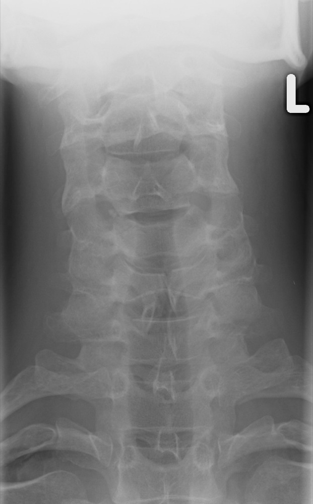
    It is important that lines 1 and 2 are traced to the superior aspect of the odontoid peg:

Normal convex lines
  • Alignment of C7 with T1 must be demonstrated. If not on the initial lateral, a swimmer's view or trauma obliques will demonstrate this area.
  • An increase in the retropharangeal soft tissues may be caused by haemorrhage or oedema due to a fracture or dislocation. The upper limit of normal is:

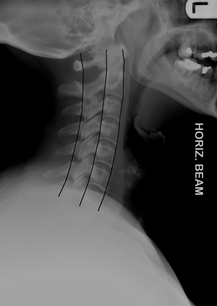
    C1 - C4 = 4-7mm (should "hug" the anterior cortex)
    C5 - C7 = 16-20mm (roughly equal to the vertebral body)

  • The vertebral bodies and intervertebral discs should be of uniform height. If the anterior height of a vertebral body is 3mm or more less than the posterior height, this is evidence of a wedge-compression fracture.
  • The distance between the posterior aspect of the arch of C1 and the anterior aspect of the odontoid peg should be no more than 3mm in adults and 5mm in children.
  • Assess Harris' ring at the base of C2, overlying the vertebral body. This should remain unbroken anteriorly, posteriorly and superiorly.
  • Beware of a physiological anomaly in children. Up to 25% of children demonstrate "pseudosubluxation" at C2/C3 - a posterior step may be seen, but this should be no more than 2mm. This is due to laxity of the ligaments:
Pseudosubluxation C2/C3
  • Although the ligaments aren't demonstrated radiographically, clues to their disruption lie in the alignment and location of the osseous structures supported by the ligaments.

AP Radiograph
  • The AP radiograph is often overlooked, however, some fractures that are not visible on the lateral radiograph will be visible on the AP. Assess superior and inferior endplates, spinous processes, lateral masses:
Normal AP
  • Spinous processes should lie in a straight line, except if they are bifid. If there is malalignment, a unilateral facet joint dislocation may be present:
Malalignment spinous processes
  • The distance between the spinous processes should be equal. No space should be 50% wider than the one immediately above or below it. If so, this is characteristic of an anterior cervical dislocation:
Widening spinous processes
Open Mouth Radiograph (C1/C2)
  • This view enables assessment of C1 and C2 (fractures of the odontoid peg, however, are often more visible on the lateral projection due to subsequent anterior / posterior displacement - see below):
Normal C1/C2
  • Lateral masses of C1 should not overhang the lateral masses of C2. If present, this is indicative of a burst fracture:
Jefferson burst fracture - C1
  • There should be symmetric space between the odontoid peg and lateral masses of C1. However, beware that normal asymmetric widening may be seen due to rotation of the patient's head:
Asymmetric widening due to rotation
  • Beware of the Mach effect - artefacts (incisors/occiput/soft tissues) overlying the base of the peg and mimicking a fracture:
Mach effect due to overlying incisor    Mach effect - repeat is normal

"Swimmer's" View
  • Careful combination of centering, positioning, selection of adequate exposure factors, and particularly collimation are all essential to produce an optimal swimmer's view:
Normal swimmer's
  • The alignment of C7 with T1 must always be assessed after a traumatic injury to the cervical spine. Not all of the T1 vertebral body needs to be demonstrated. However, the anterior vertebral line or posterior vertebral line must be visualised in order to answer the question "is there a subluxation present?":
Unilateral facet dislocation - nondiagnostic    Unilateral facet dislocation - repeat - diagnostic

Oblique Views
  • 45° obliques will demonstrate:

    Intervertebral foramina, and the presence of osteophyte encroachment in spondylosis (OA).
    Facet joints.
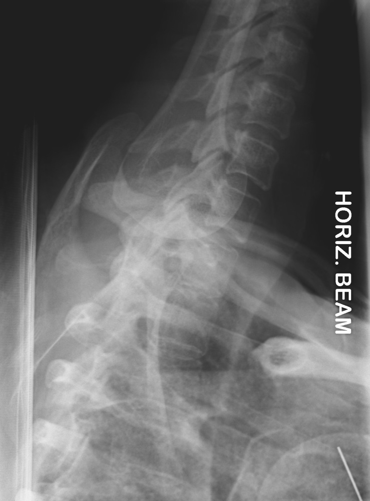
    Alignment of C7 with T1 if the swimmer's view is unobtainable (30% obliques satisfactory).

  • Right posterior oblique demonstrates the left foramina.
  • Right anterior oblique demonstrates the right foramina.
Facet joint dislocation - right oblique    Facet joint dislocation - left oblique    Facet joint dislocation - lateral

Flexion and Extension Views
Normal flexion   Normal extension
  • To demonstrate ligament instability and subsequent vertebral mobility.
  • Radiographs are often taken on patients with rheumatoid arthritis prior to having a general anaesthetic, as RA causes the ligaments to become lax:
RA with atlanto-axial subluxation in flexion    RA with normal alignment in extension
  • In the context of trauma, flexion and extension views are taken when there is mild mal-alignment on plain radiograph and index of suspicion is high for ligament rupture.
Soft Tissue Structures
Main Ligaments
  • Anterior Longitudinal Ligament
  • This is a taut, strong structure that is closely applied to the anterior aspects of the vertebral bodies and the annulus of the intervertebral discs.

  • Posterior Longitudinal Ligament
  • This is weaker, and is attached to the posterior vertebral bodies and intervertebral discs.

  • Ligamentum Flavum
  • This ligament lines the dorsal surface of the spinal canal and is tightly applied to the laminae.

  • Interspinous Ligaments
  • These interconnect the spinous processes.

  • Supraspinous Ligament
  • Is applied to the dorsal tip of the spinous processes, and overlies the interspinous ligaments.

Intervertebral Discs
  • The intervertebral discs consist of:
    • A central, gelatinous nucleus pulposus.
    • Surrounding peripheral, concentric layers of annulus fibrosis.
Spinal Canal
  • The normal anteroposterior diameter of the cervical spinal canal is approximately 10-20mm. On the lateral radiograph it may be measured relative to the vertebral body - the two should be equal. In the cervical spine region, the spinal cord occupies 50% of the spinal canal.
Pathomechanics of Cervical Spine Trauma
Flexion
  • Up to 80% of all cervical spine injuries are due to hyperflexion.
  • As the head is flexed, the maximum force is focused upon the bodies of C4-C7.
  • Compression of the vertebral body causes anterior wedging.
  • The posterior elements - the spinous processes, laminae and supporting ligaments are placed in tension, which result in fractures and tears of these structures.

Extension
  • Hyperextension creates tension in the anterior longitudinal ligament. This may tear at the intervertebral disc space or at the margin of the vertebral body. The latter results in an avulsion fracture of the anterior superior or inferior margin of the vertebral body.
  • The posterior elements are simultaneously compressed, which may result in fractures of the spinous processes, laminae and facets.

Axial Compression
  • Initial trauma is to the vertebral endplates.
  • Increased compression causes the intervertebral disc to explode into the vertebral body. This creates a comminuted fracture.

Distraction
  • Movement of the head, which weighs approximately 4-5kg, creates tensile forces on the cervical spine.
  • If occurring in combination with flexion or extension, this lessens the severity of compression resulting from these forces. Osseous structure will be maintained, to the detriment of the interspinous ligaments.

Rotation
  • The spinal ligaments withstand compression and distraction forces well, but are very susceptible to disruption by rotation.
  • Results also in fractures of the posterior elements, particularly the facets and laminae, and in fracture - dislocations.
Significance of Injury and the concept of stability
  • The spine may be split into three "columns" for the purpose of assessment of stability:
    1. Anterior column - Involves the anterior two thirds of the vertebral body/intervertebral disc, and the anterior longitudinal ligament.
    2. Middle column - Involves the posterior aspect of the vertebral body/intervertebral disc, and the posterior longitudinal ligament.
    3. Posterior column - Involves the posterior elements - the lamina, facet joints, spinous processes, and the associated ligaments.
  • An injury to the spine is considered unstable if two of the three columns are disrupted. Generally, if the middle column is disrupted, either the anterior or posterior columns are also involved, and the injury is unstable.
  • The middle column is the fulcrum from which the spine pivots into flexion and extension. It is generally thought that the middle column remains intact, and is therefore stable, in simple flexion and extension injuries. Axial compression, distraction and rotational injuries, or a combination of these with flexion or extension, usually disrupt the middle column.
Cervical Spine Trauma
Atlas (C1)
  • Neural arch fracture
  • This is a longitudinal fracture through the posterior neural arch, usually bilateral. It is caused by hyperextension, with the result that the neural arch of C1 is compressed between the occiput and C2. It is best demonstrated on the lateral projection:

Neural arch fracture
  • Burst (Jefferson) fracture
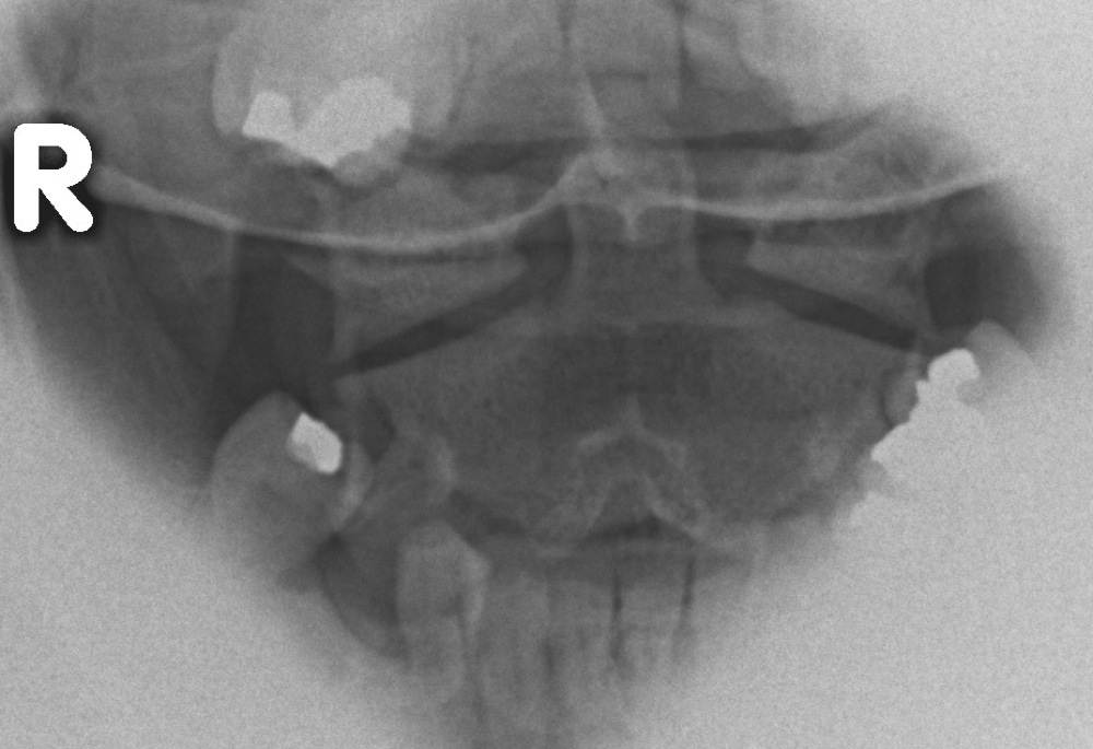
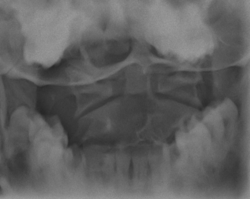
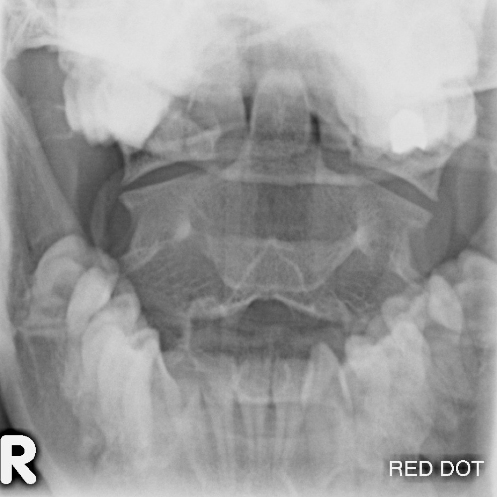
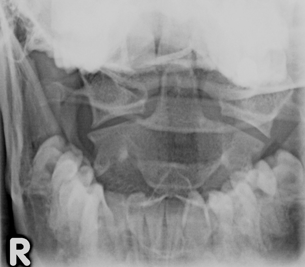
  • This is a comminuted fracture, with bilateral disruption of both anterior and posterior arches, and lateral displacement of both lateral masses. It is caused by axial compression with the transmission of force from the skull downwards through the occipital condyles, compressing the lateral masses. Demonstrated on the open mouth view by:

    • unilateral C1/C2 odontoid peg joint space widening.
    • lateral masses of C1 overhang the lateral masses of C2.
    •  

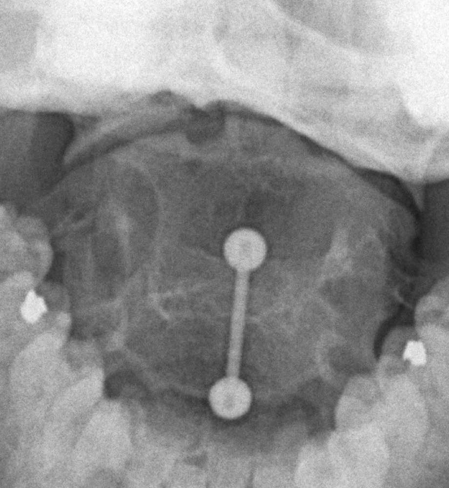
    The injury is considered stable if the overhang is less than 7mm. Over 7mm indicates that the transverse ligament is disrupted, and is therefore unstable:

Jefferson burst fracture - C1
Axis (C2)
  • Odontoid peg fracture
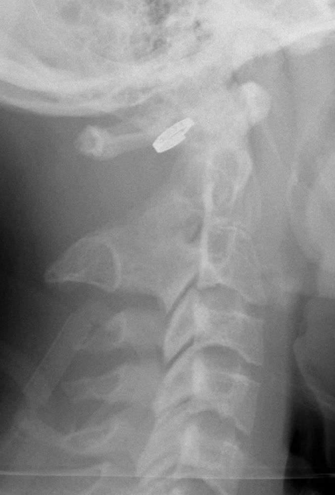
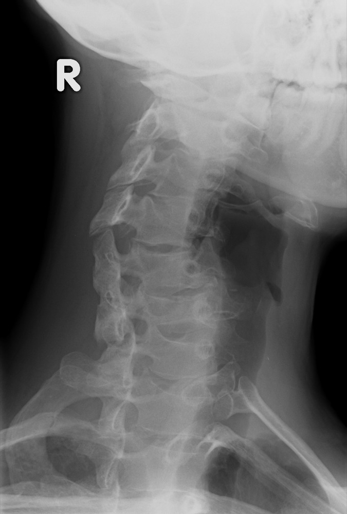
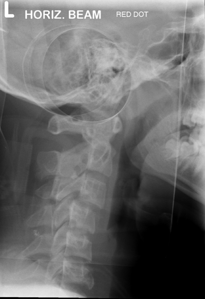
  • This is the most common fracture of C2. May be caused by flexion or extension and usually results in ligamentous instability. It usually involves the base of the peg and may be visualised on either the open mouth or, more commonly, lateral view. Assess for any soft tissue swelling anteriorly. Also look carefully at Harris' ring on the lateral projection:

Odontoid peg fracture    Odontoid peg fracture    Odontoid peg fracture
  • Hangman's fracture (traumatic spondylolisthesis)
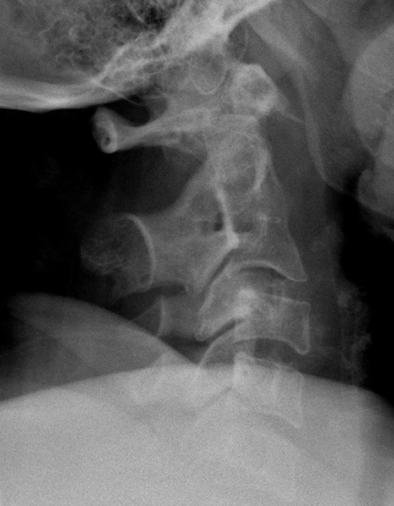
  • Hyperextension of the neck transmits the force through to the C2 pedicles. This results in an oblique fracture originating anterior to the inferior facet of C2 and extending supero-posteriorly. Tension causes disruption of the anterior longitudinal ligament causing this injury to be unstable. Will be demonstrated on the lateral view but may be undisplaced:

Hangman's fracture - C2
C3-C7
  • Anterior wedge compression fracture
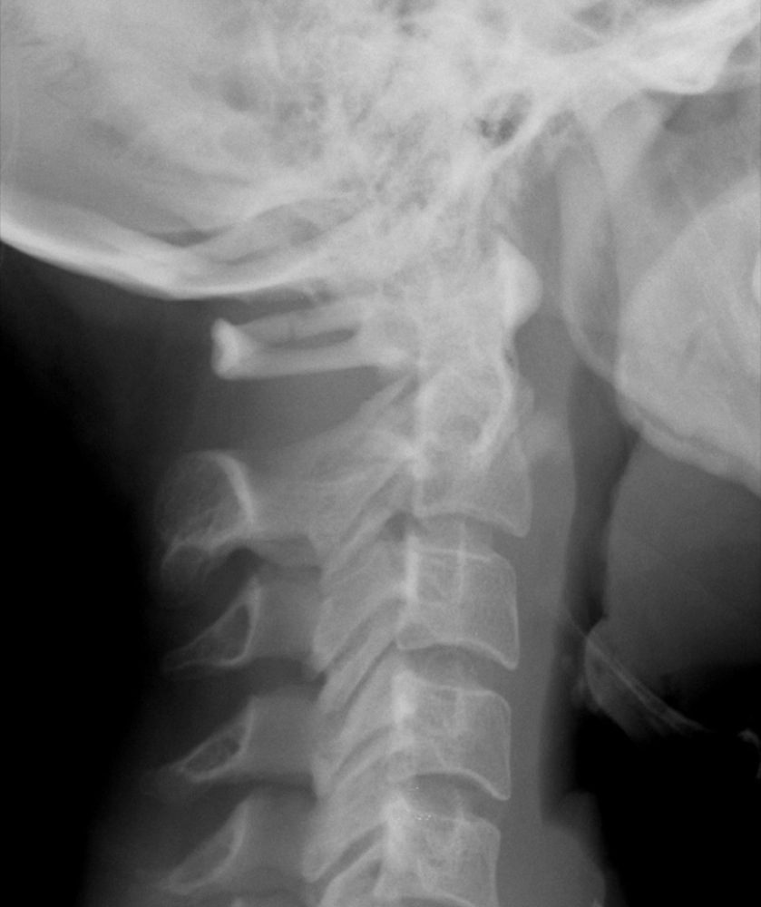
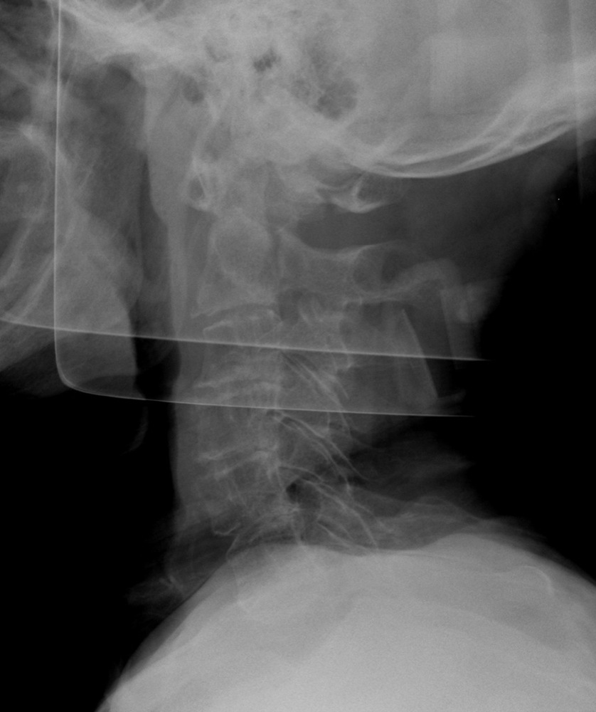
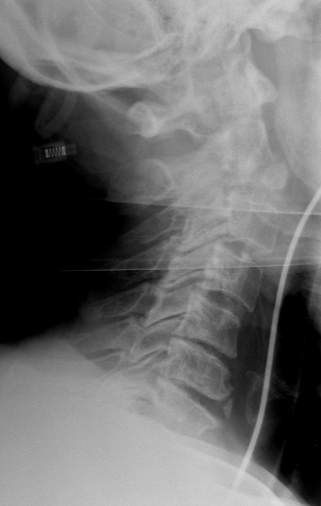
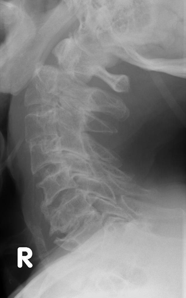
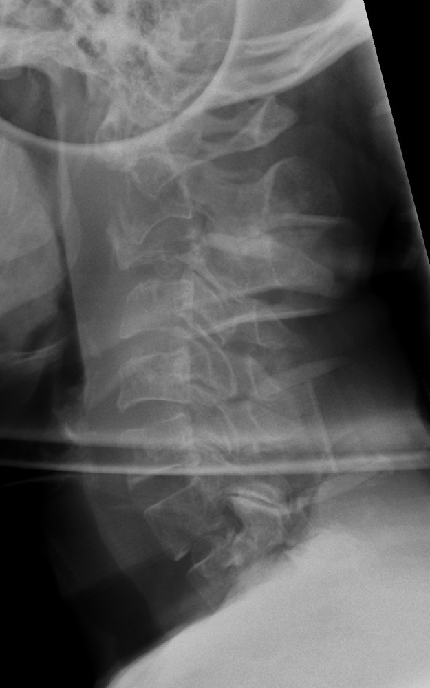
  • This type of fracture is caused by hyperflexion with the result that the vertical height of the vertebral body is decreased anteriorly, as viewed on the lateral radiograph. The posterior elements remain intact. This is a stable injury:

Anterior wedge compression fracture - C7
  • Burst fracture
  • Caused by axial compression, the intervertebral disc is driven into the vertebral body below. The vertebral body explodes into several fragments; a fragment from the postero-superior surface being driven posteriorly into the spinal canal. This is an unstable injury that frequently results in spinal cord injury. It is therefore important to check the posterior vertebral cortex for evidence of disruption, on an apparently simple wedge compression injury.

  • Unilateral locked facet
  • Flexion, rotation and distraction may cause the facet joints on one side to be locked. This results in the vertebra being displaced anteriorly by 25%, as demonstrated on the lateral radiograph. The facet joints are seen in true lateral profile above and oblique profile below, or vice versa:

Unilateral perched facet - C3/C4
  • Bilateral locked facets
  • If the amount of distraction increases, the facets may become disarticulated. The vertebral body is displaced anteriorly by 50%, and the inferior facets of the anteriorly displaced vertebra lie anterior to the superior facets of the vertebra below. Assess both anterior and posterior vertebral lines and also look carefully at the facet joints; they should have a roof tile appearance, parallel to one another:
Bilateral perched facets - C5/C6
  • Teardrop extension fracture
  • Hyperextension causes a triangular fragment to be avulsed off the antero-inferior corner of the vertebral body. This is not associated with any neurological damage. The axis is most commonly involved:

Teardrop extension fracture - C2
  • Spinous process fracture
  • This is an avulsion by the supraspinatous ligament off the spinous process, usually C6 or C7. This is caused by flexion as the body rotates relative to the head and neck. Usually undisplaced and therefore only seen on the lateral radiograph:

Spinous process fracture - C4
  • Whiplash
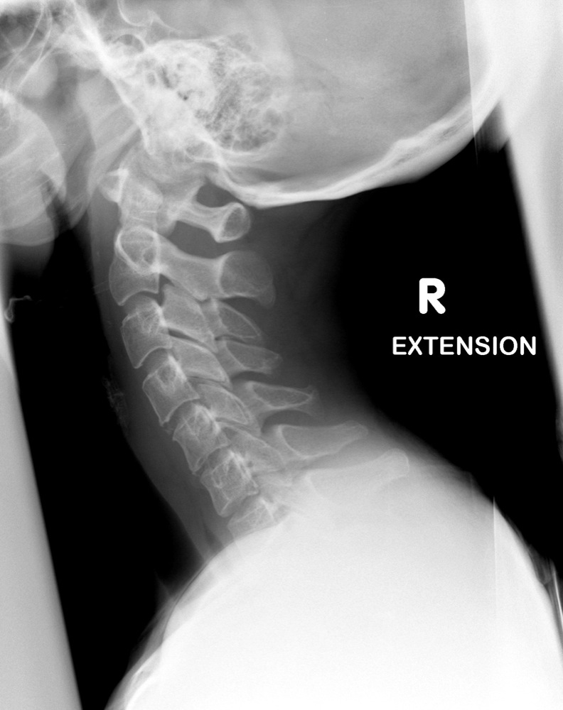
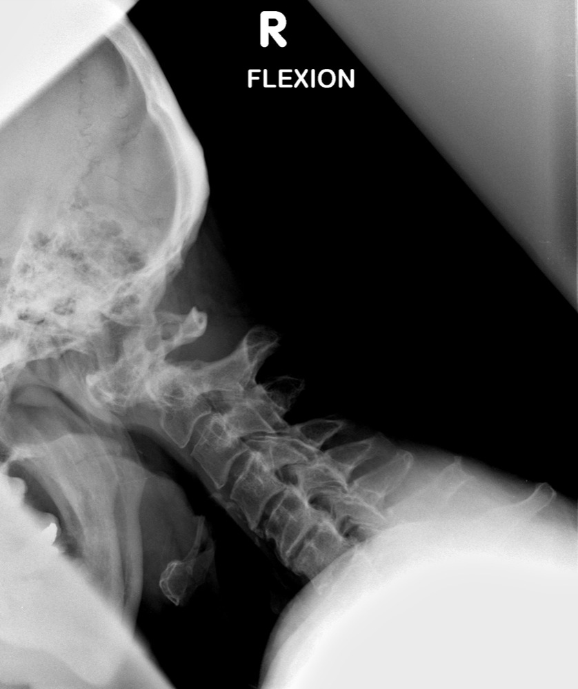
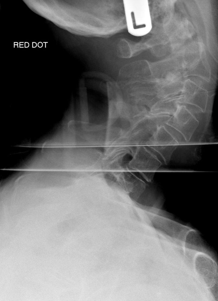
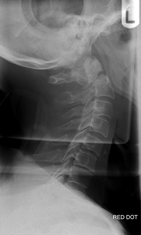
  • Sudden deceleration of the body, with flexion and extension movements of the cervical spine usually results in sprain or intervertebral disc injury without fracture or dislocation. The commonest radiographic appearance is straightening of the cervical spine due to severe muscle spasm, with the normal curvature reduced or reversed:

Whiplash - reversal of the normal lordosis
  • Hyperflexion Strain
  • Refers to soft tissue injury. Anterior subluxation occurs with disruption of the posterior longitudinal ligament, the interspinous ligament and the intervertebral disc. The lateral projection demonstrates localised kyphotic angulation with an increase in height of the intervertebral disc posteriorly and associated fanning of the spinous processes.

  • Hyperextension Strain
  • The converse of hyperflexion strain; the anterior longitudinal ligament is disrupted as evidenced by widening of the intervertebral disc space anteriorly. The facet joints are disrupted and the interspinous distance is narrowed.

Pathology
  • Spondylosis
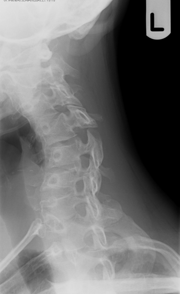
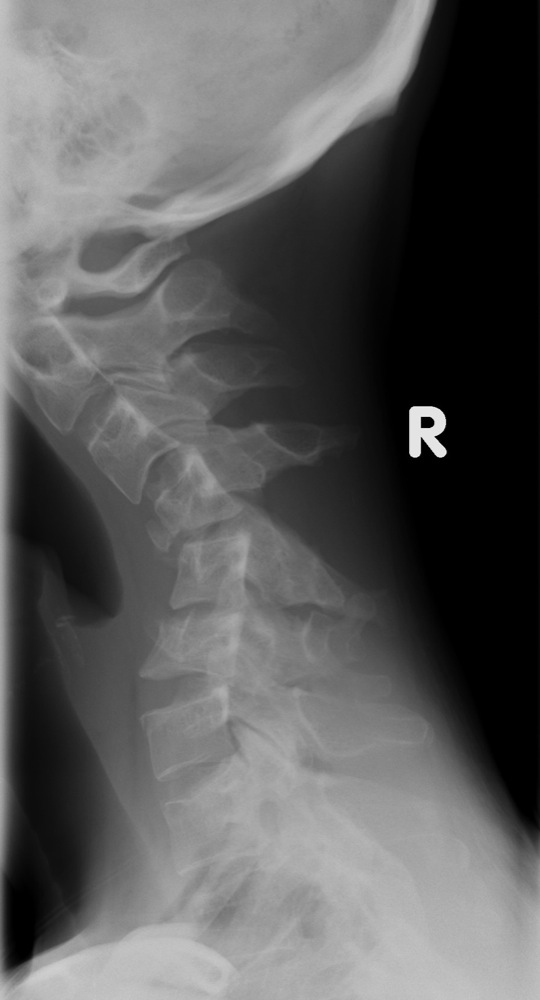
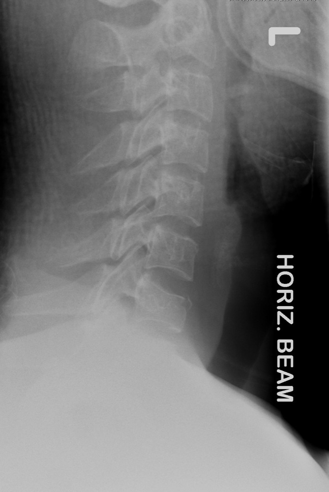
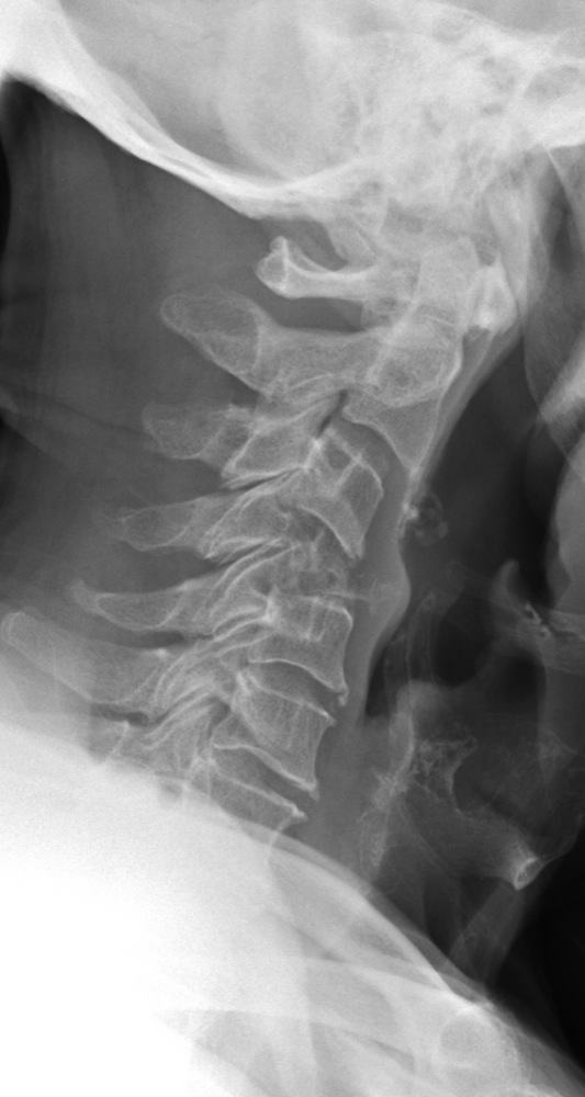
  • Refers to degenerative changes of the intervertebral disc spaces, which is demonstrated by disc space narrowing, endplate sclerosis and osteophyte formation. Facet joint OA is seen posteriorly. The associated osteophytes may impinge on the nerve root foramina. The appearance of striking degenerative changes within the cervical spine may obscure underlying injury. It is therefore important to search for co-existant trauma. A common mechanism of injury in those patients with spondylosis (often the elderly) is a fall directly onto the forehead with a subsequent fracture at C2:

Fracture C2 with spondylosis    Fracture C2 with spondylosis    Fracture C2 with spondylosis
  • Metastatic disease
  • Primary tumours can metastasise to the vertebral bodies demonstrating a lucent, moth-eaten, permeative appearance. There is often subsequent collapse:

Metastatic disease C2,C3    Metastatic disease C4
  • Congenital fusion
  • It is not unusual to see a congenital fusion within the cervical spine, usually at C2/C3 with fusion of the vertebral bodies and posterior elements. This is often associated with a hypoplastic odontoid peg:

Congenital fusion C2,C3    Congenital fusion C2,C3
Congenital fusion, hypoplastic peg    Congenital fusion, hypoplastic peg

TAKE THE CERVICAL SPINE SELF-TEST

- return to top -