Norwich Image Interpretation Course

Heidi Nunn (Advanced Practice Reporting Radiographer)


The Facial Bones

Facial bones projections Three lines Orbital fractures Zygomatic fractures Maxilla fractures Mandibular projections Anatomy of the mandible Mandibular fractures

(hover over images to zoom, click to enlarge)

Learning outcomes
  • Assess skeletal radiographs using a systematic approach
  • Use common lines which are helpful in interpreting the image
  • Understand the different radiographic projections and how the anatomy changes with position
  • Describe facial anatomy
  • Understand mechanisms of injury and the likely fractures which may result
  • Search for subtle injuries and understand their clinical significance
  • Recognise potential soft tissue injuries
Facial Bones Projections
Occipito-mental View
  • The PA occipito-mental view provides excellent demonstration of the upper and middle thirds of the face including the orbital margins, frontal sinuses, zygomatic arches and maxillary antra:

Normal facial bones
Occipito-mental 30 View
  • With 30 degrees of caudal angulation, the orbits are demonstrated less well. However, the zygomatic arches and the walls of the maxillary antra are seen clearly:

Normal facial bones
Three lines for inspecting the OM views
Normal lines    Normal lines
Line 1:
  • Look for widening of the zygomatico-frontal sutures
  • Fractures of the superior rim of the orbits
  • "Black-Eyebrow" sign due to orbital emphysema
  • Opacification / air-fluid level in the frontal sinuses
Line 2:
  • Look for fractures of the superior aspect of the zygomatic arch
  • Fractures of the inferior rim of the orbits
  • Soft tissue shadow in the superior maxillary antrum
  • Fractures of the nasoethmoid bones and medial orbits
Line 3:
  • Look for fractures of the inferior aspect of the zygomatic arch
  • Fractures of the lateral maxillary antrum
  • Opacification / air-fluid level in the maxillary sinuses
  • Fractures of the alveolar ridge
Compare the injured side with the uninjured side.

Fractures of the Facial Skeleton
  • Within the facial skeleton, there are relative areas of strength, which tend to be spared by fractures lines. These are:
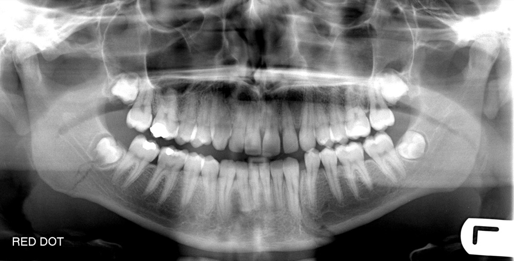
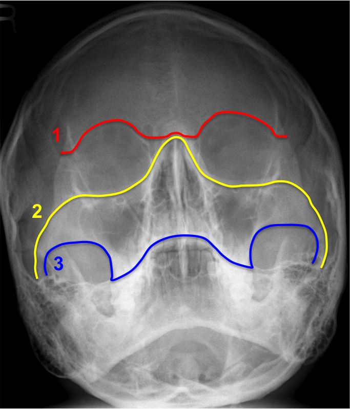
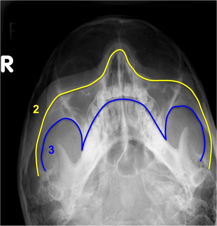
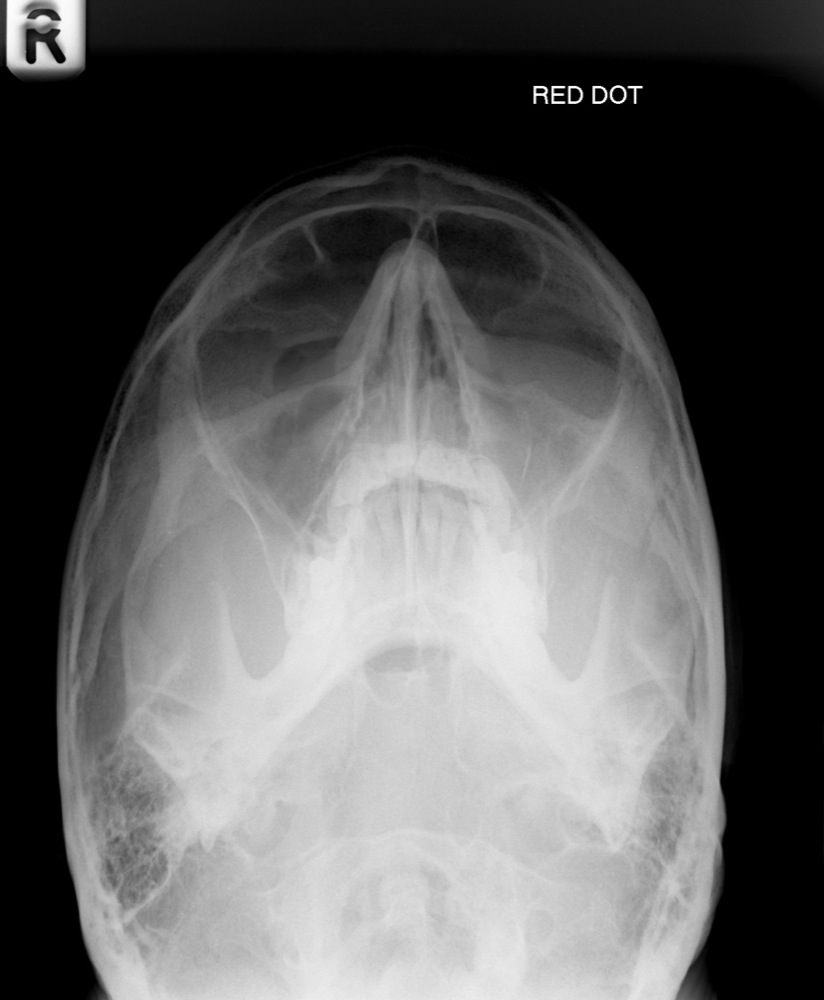
  • Alveolar ridge of the maxilla
    Nasofrontal process of the maxilla
    Body of the zygoma

Fractures of the Orbits
  • Blowout fracture

    The most common portion of the orbit to sustain a fracture is the weak floor, and this injury, if occurring in isolation, may result in a blowout fracture. A blow to the globe causes increased intraorbital pressure. This causes a fracture of the thin plate of bone forming the floor of the orbit (i.e., the roof of the maxillary antrum). Some of the orbital contents such as fat and muscle herniate downwards into the maxillary sinus resulting radiographically in a soft tissue "teardrop", or polypoid mass, in the roof of the maxillary antrum:

Blowout fracture - left
  • Orbital emphysema

    Herniation through the medial wall may also occur. Air from the maxillary or ethmoid sinuses may enter the orbit, giving rise to orbital emphysema. This is commonly known as the "black eyebrow" sign:

Orbital emphysema - right    Orbital emphysema - right
Fractures of the Zygoma
  • Tripod fracture

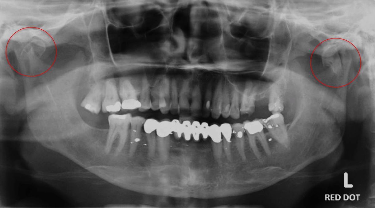
    The zygomatic bone has three distinct limbs; the orbital process leading to the zygomatico-frontal suture, the zygomatic arch and the maxillary process surrounding the superior and lateral margins of the maxillary antra. Trauma to the facial skeleton tends to spare areas of relative strength, and the body of the zygoma is one such area. It therefore follows that fractures are likely to be sustained at these three limbs and such an injury is termed the tripod fracture.

  • Radiographically, this common injury manifests itself as:

    • Widening of the zygomatico-frontal suture.
    • Fracture of the zygomatic arch.
    • Fracture of the inferior orbital rim extending through the anterior and lateral walls of the maxillary antrum.
Tripod fracture - left    Tripod fracture - left
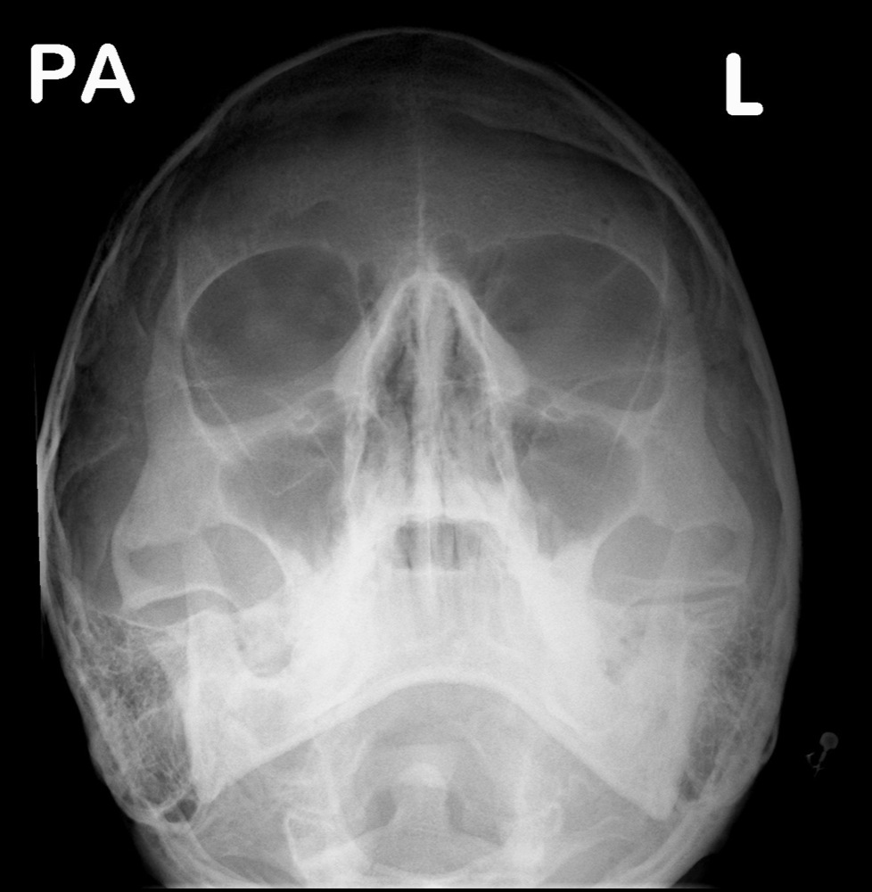
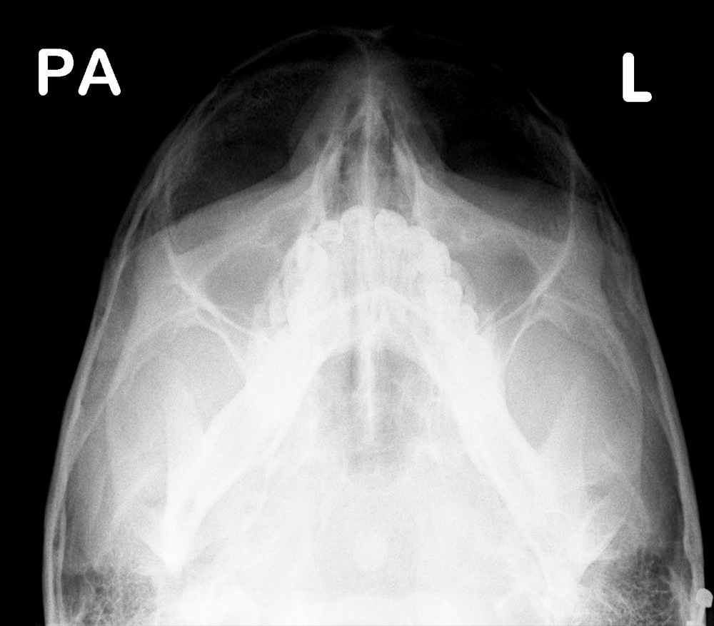
  • Both the OM and OM 30 views will identify these fractures, along with the associated soft tissue swelling over the zygomatic eminence and opacification or an air-fluid level in the maxillary sinus. Often only a straight PA would identify the diastasis at the zygomatico-frontal suture.

  • Isolated fractures of the zygomatic arch

    These generally consist of three fracture lines with medial and inferior displacement of the fragments. More often than not, however, an apparently isolated fracture of the zygomatic arch is actually part of a more complex tripod fracture.

Zygomatic arch fracture - left
Fractures of the Maxilla

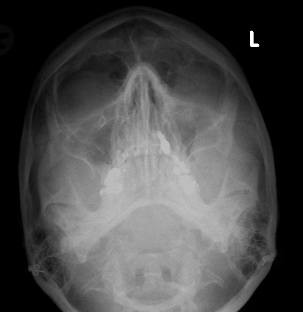
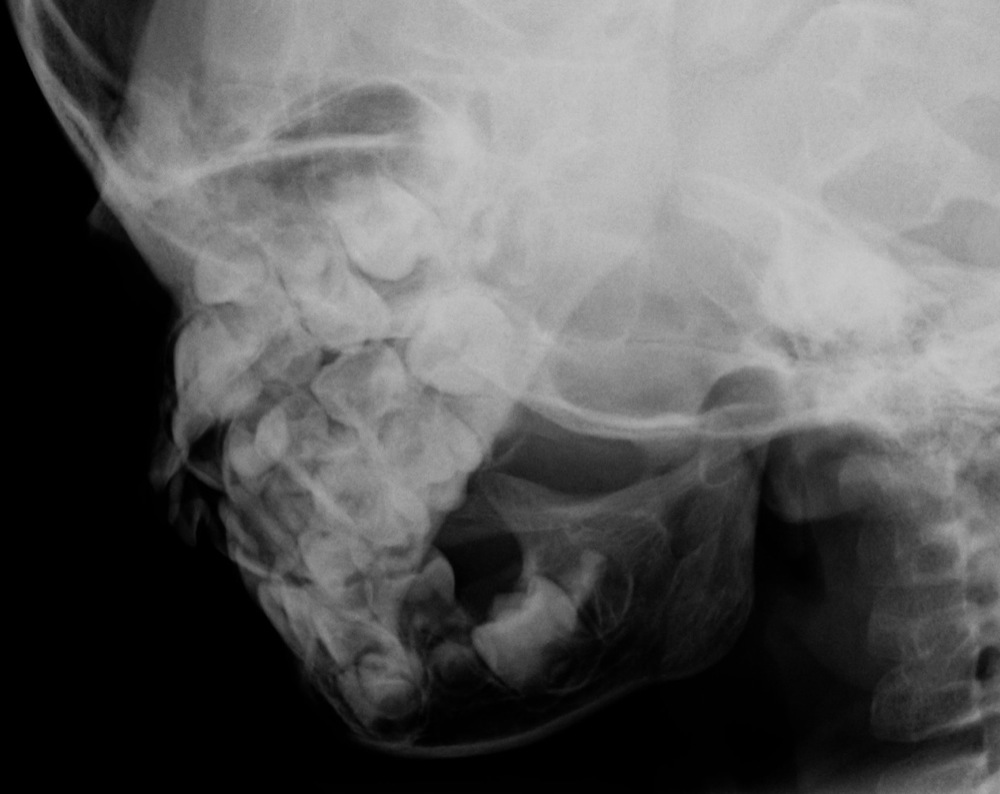
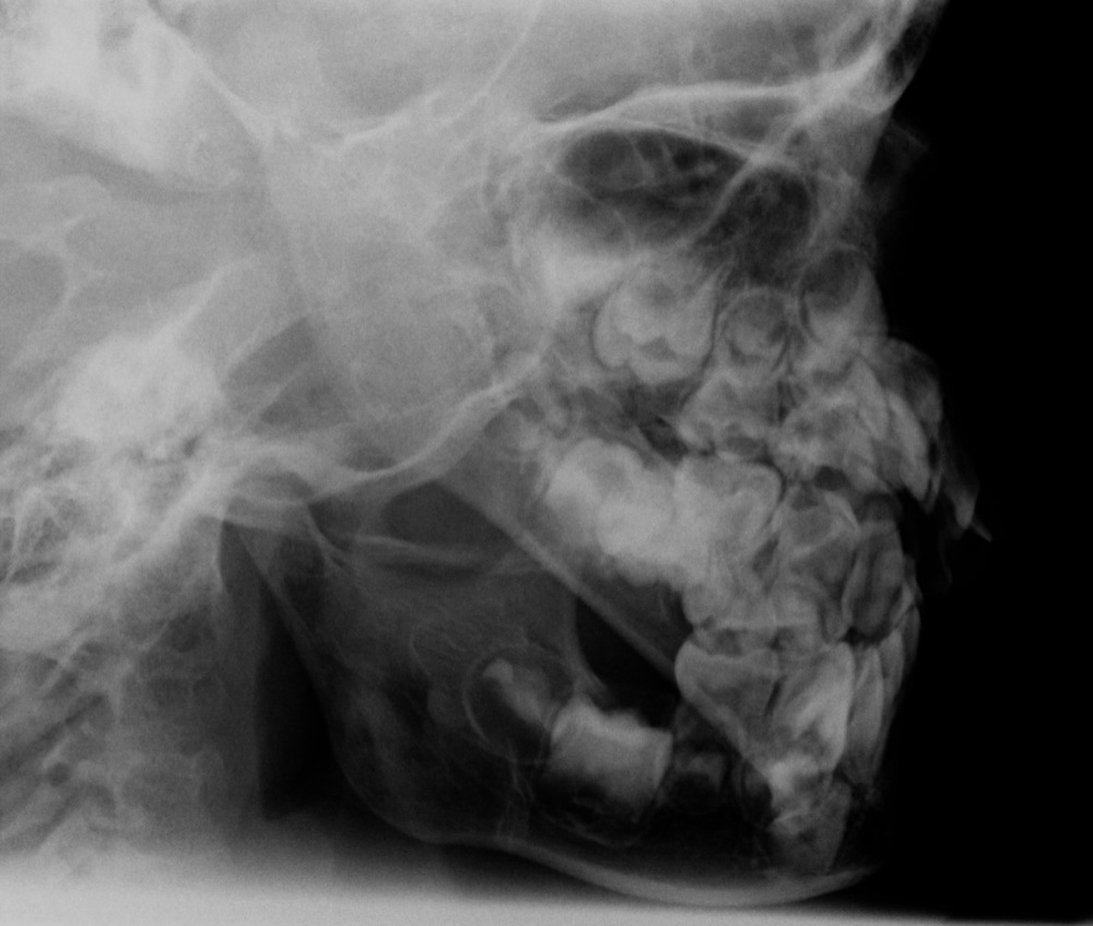
The classification of maxilla fractures again follows the concept of areas of relative strength within the facial skeleton. There are three principal fracture lines which correspond to relative areas of weakness, and these are referred to as Le Fort fractures. By definition, these fractures must transect the pterygoid process of the sphenoid bone.

Le Fort I

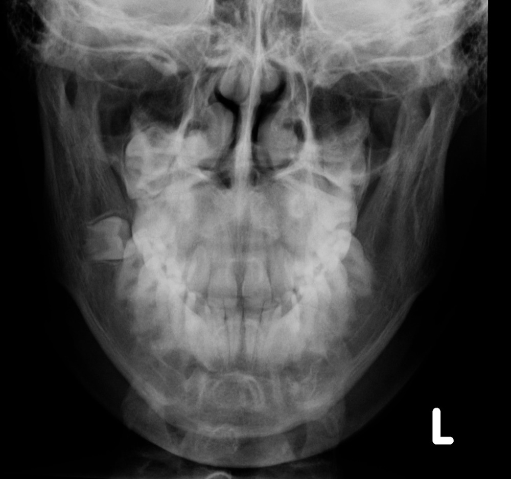
This is a transverse fracture through the inferior maxillary antra, which separates off the alveolar process of the maxilla. The Le Fort I is demonstrated on the OM view with fractures through the medial and lateral walls of the maxillary antra, and the nasal septum.

Le Fort II

This is a pyramidal fracture, which separates off the central portion of the face. The OM radiograph identifies the Le Fort II with fractures through the lacrimal bones, medial orbital walls, infra-orbital rim and lateral walls of the maxillary antra.

Le Fort III

This fracture is characterised by separation of the entire facial skeleton from the skull. The posterior aspect of the fracture extends down the posterior maxillary sinus walls. Fracture lines will be visible on the OM view extending from the medial orbits and nasoethmoid region across the ethmoids posteriorly. The orbits appear elongated with wide diastasis of the zygomatico-frontal sutures, or fractures of the orbital process of the zygoma.

In practice, this classification is over-simplified, and in reality, fractures tend to be more complex. There may be a combination of appearances, such as a Le Fort II on one side, and a Le Fort III on the other. Also, as these injuries are associated with major trauma, these patients tend to go direct to CT and plain radiograph examples of classic Le Fort fractures are infrequent.
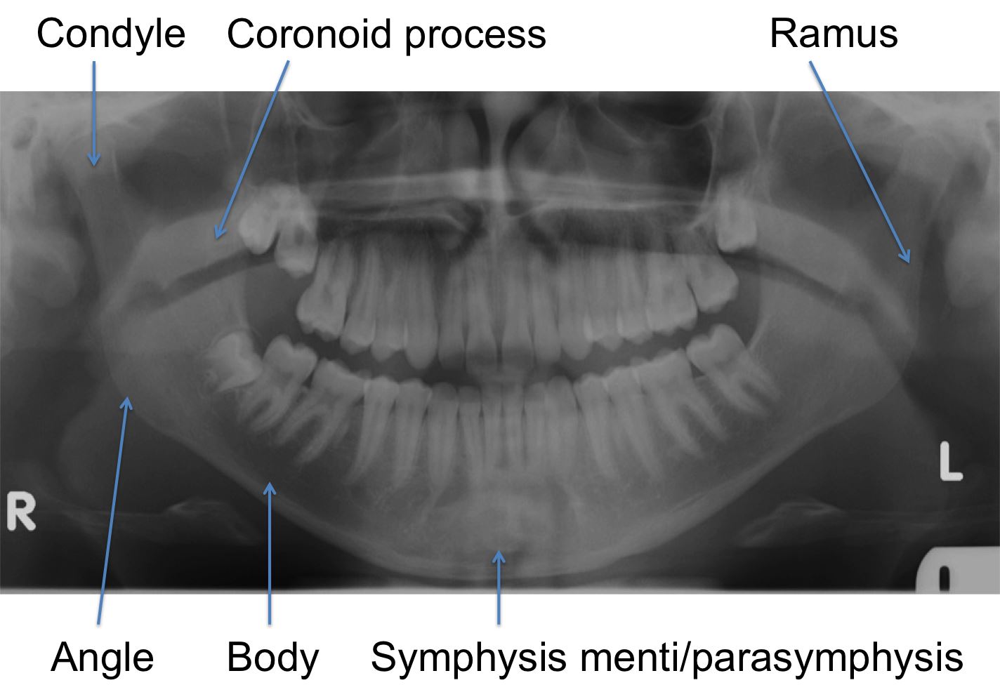
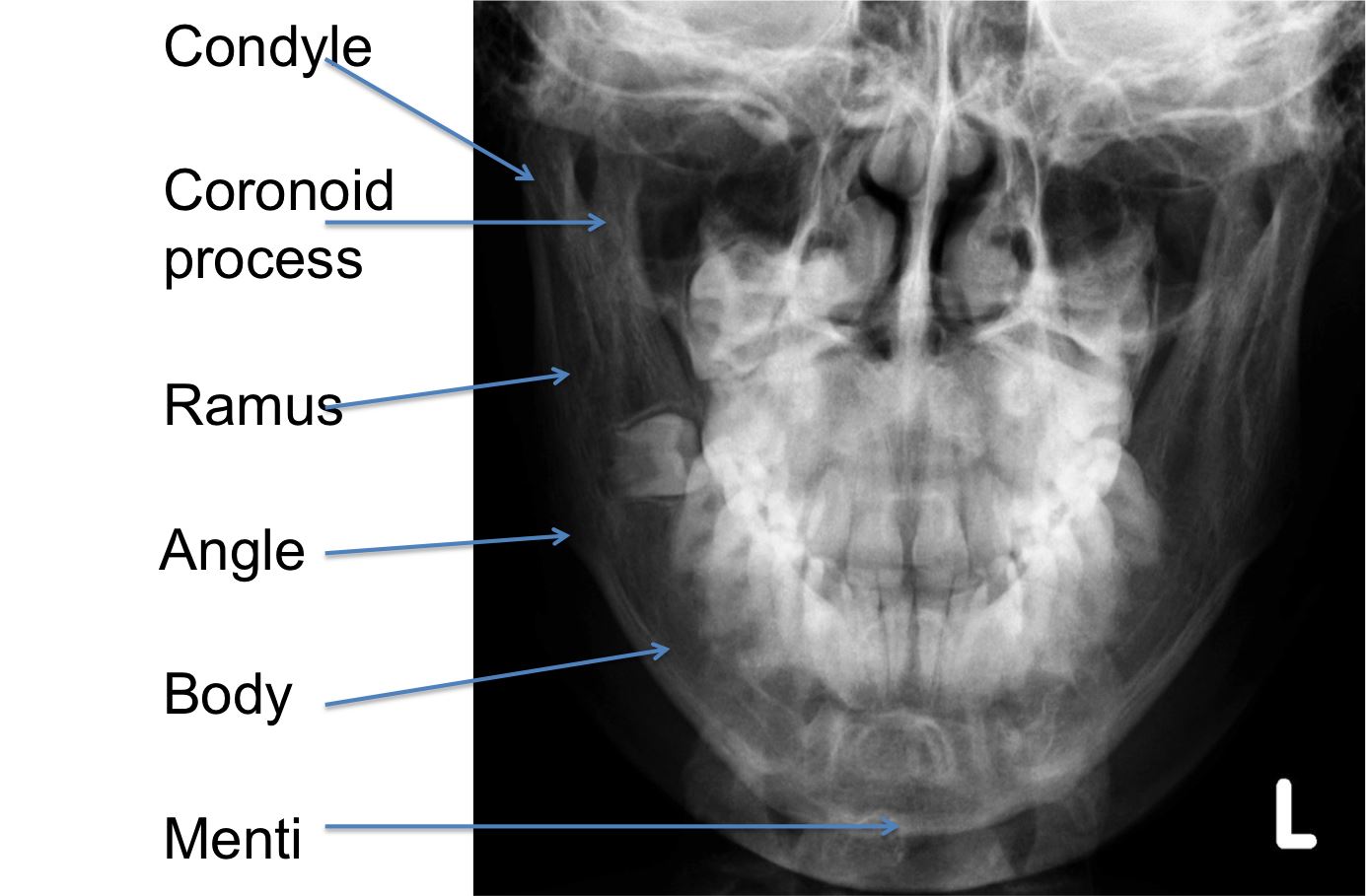
Mandibular Projections
Orthopantomogram (OPG)
  • This is an excellent method of demonstrating virtually all mandibular fractures, including the coronoid and condylar processes. Be aware however; occasionally symphysis menti fractures may not be seen.
Normal OPG

Posteroanterior (PA) Mandible
  • The OPG must be supplemented with a PA mandible to establish displacement of fractures. This will demonstrate the body and symphysis menti, and also provide tangential views of the rami and the necks of the condyles:
Normal PA mandible
Lateral obliques
  • In the absence of an OPG, the lateral obliques will demonstrate the body and ramus on each side. The articulation at the temporomandibular joint may also be assessed:
Normal lateral oblique - paediatric    Normal lateral oblique - paediatric
Anatomy of the mandible
Mandibular anatomy    Mandibular anatomy
Fractures of the mandible
  • The distribution of fractures of the mandible are:
Condyle
15-20%
Coronoid process
1-2%
Ramus
3-9%
Angle
20-31%
Body
21-40%
Symphysis menti
10-15%
Alveolar ridge
3-4%

  • Fractures of the mandible tend to occur in more than one place, therefore once one has been identified, another should be sought. The second injury may be a fracture, or a temporomandibular joint dislocation.
  • The mandible is composed of an inner and outer cortex enclosing the medullary cavity. Fracture lines may be identified at the both inner and outer cortices but this represents one fracture:
Fracture right angle and left body
  • Fractures of the coronoid and condylar processes often occur at their bases and may be difficult to visualise:
Bilateral condyle fractures
  • The PA mandible will demonstrate medial (or less commonly lateral) angulation of the condyle by the pterygoid muscle:
Fracture right body, left coronoid and ramus    Fracture right body, left coronoid and ramus
  • Air in the pharynx may also be identified on the OPG overlying the condyle and ramus. This should not be mistaken for a fracture.
  • Fractures are classified as either favourable or unfavourable. A favourable fracture is one that is held in apposition and alignment to the natural pull of the attached muscles. Unfavourable fractures are those that are displaced by the pull of attached muscles.
  • After diagnosing a fracture, it is important to identify any pathological dental condition in which the fracture lies. This is best demonstrated on the OPG. A fracture that extends into a periapical abscess may result in delayed healing and osteomyelitis.

TAKE THE FACIAL BONES SELF-TEST

- return to top -