|
Under 10 years
|
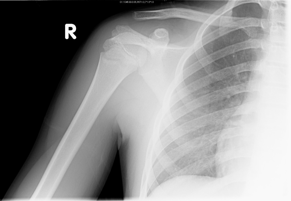
Fractured clavicle |
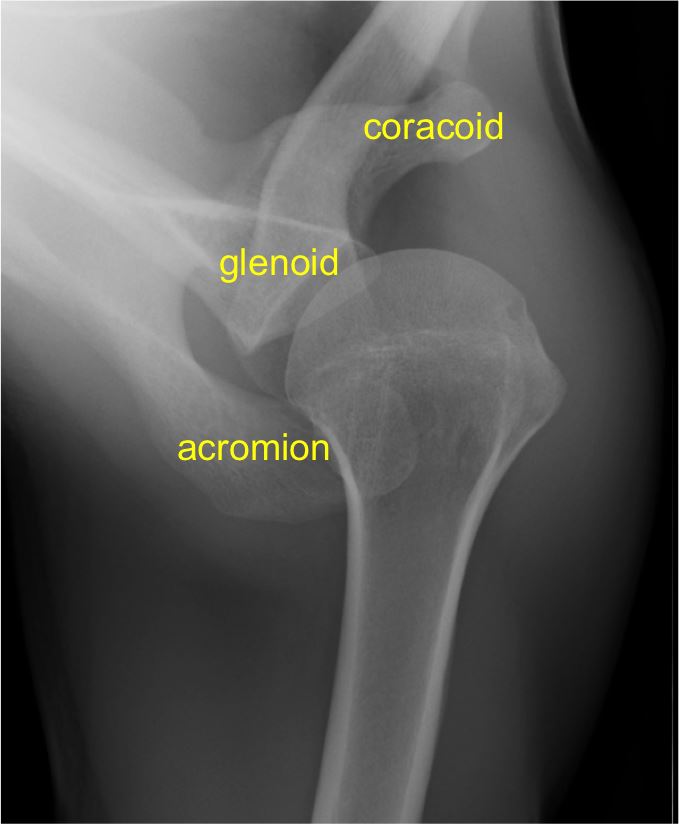
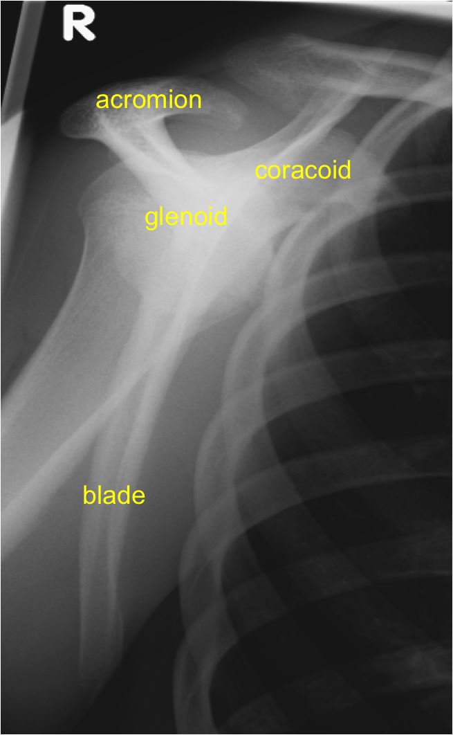
| 15-40 years | Acromioclavicular joint subluxation |
| Glenohumeral joint dislocation | |
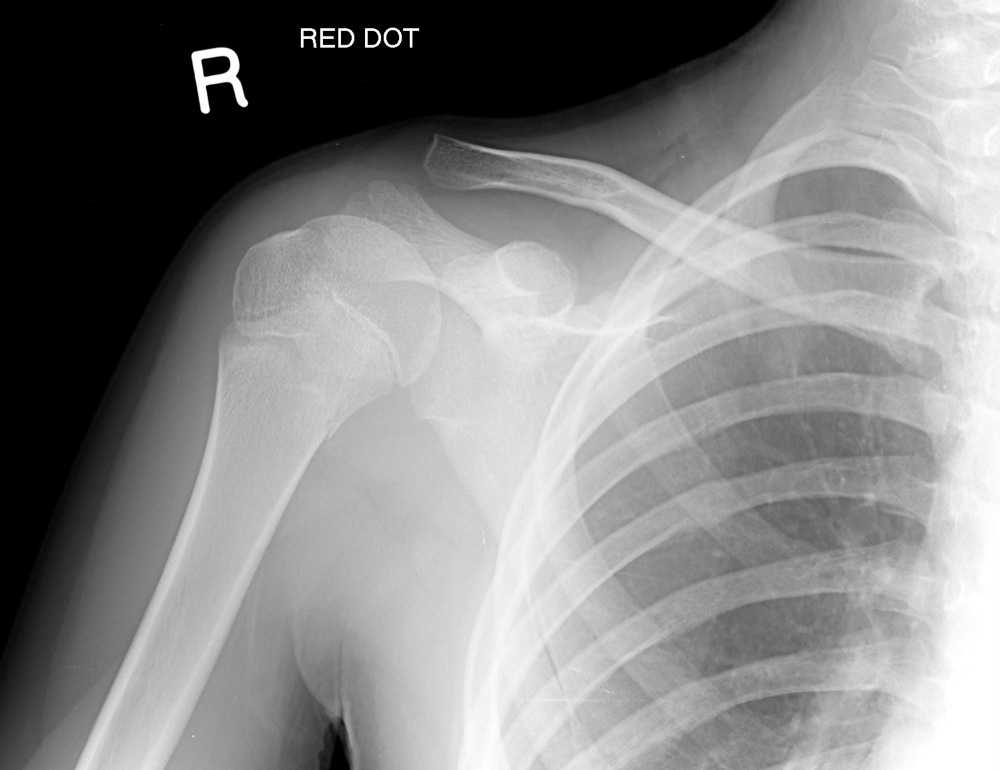
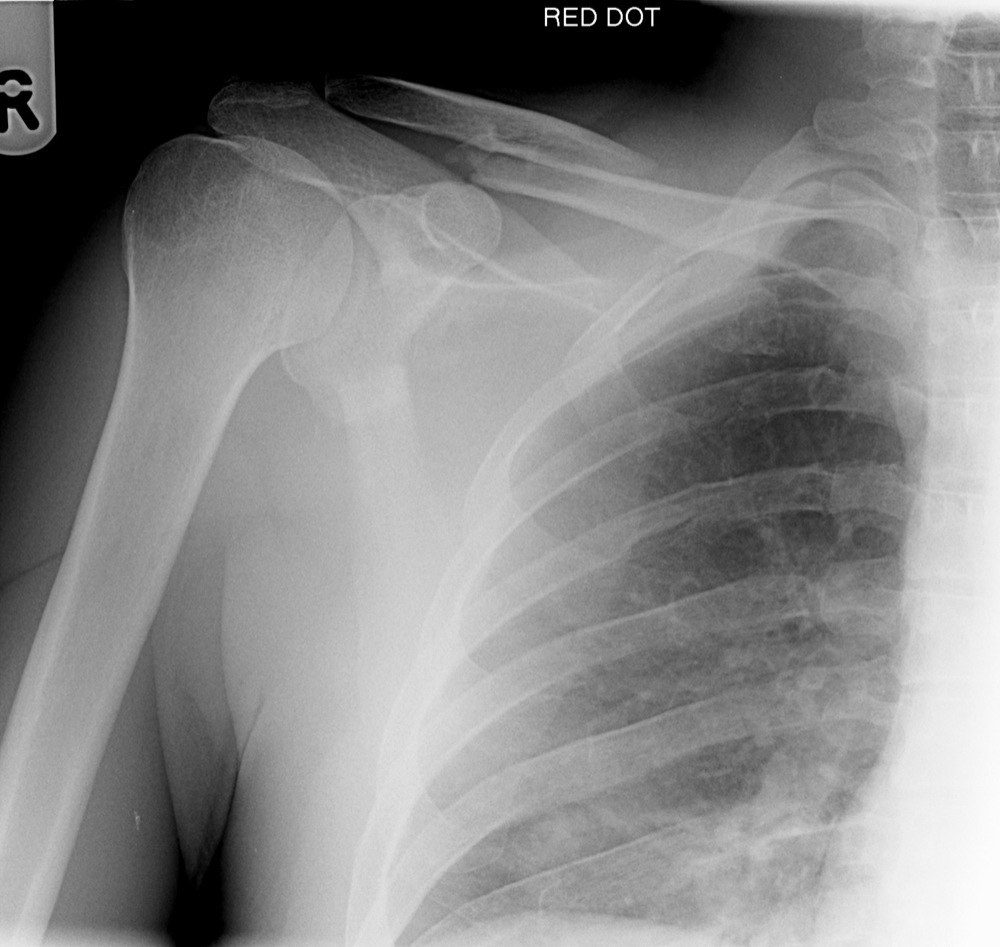
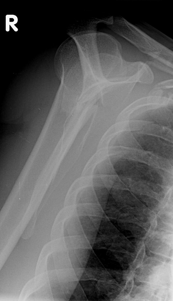
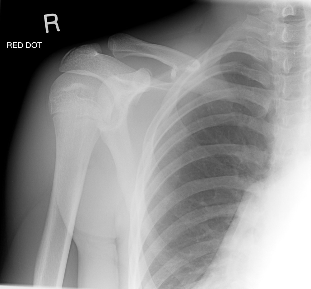
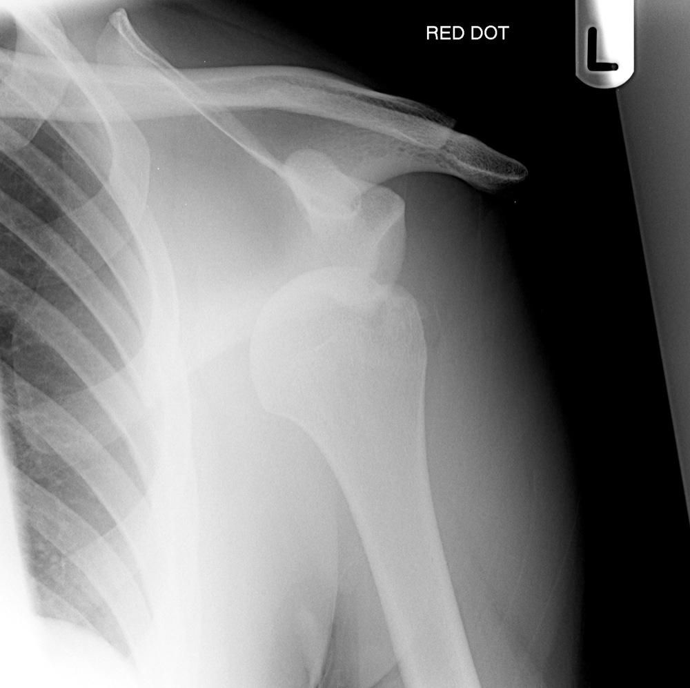
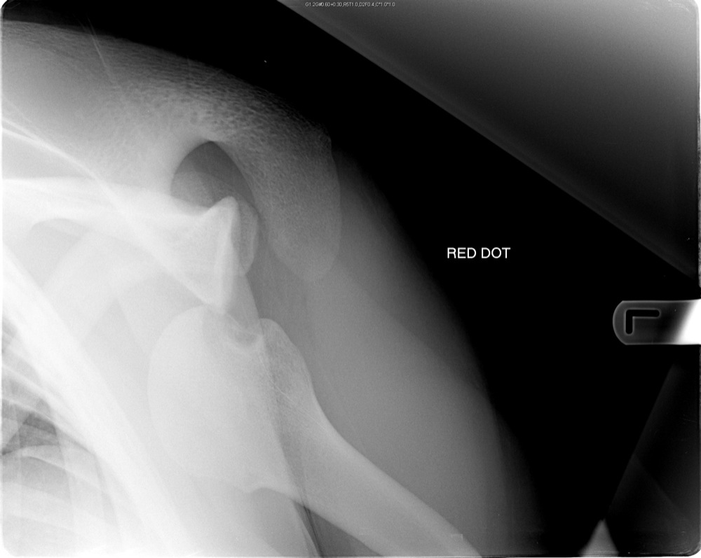
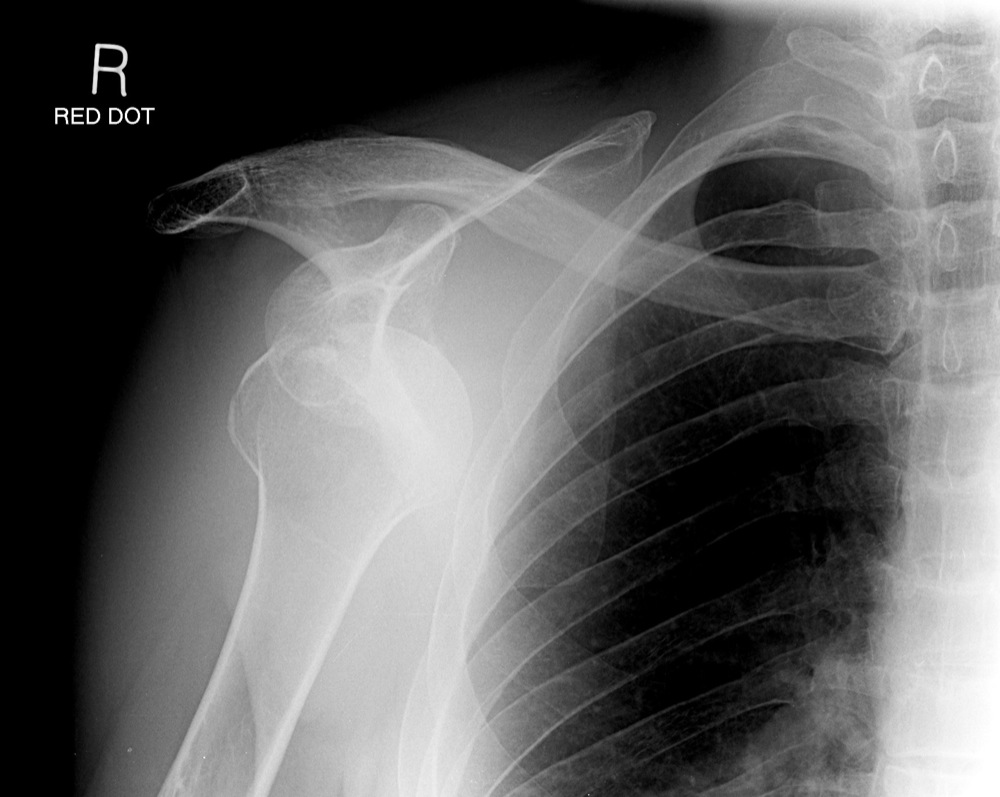
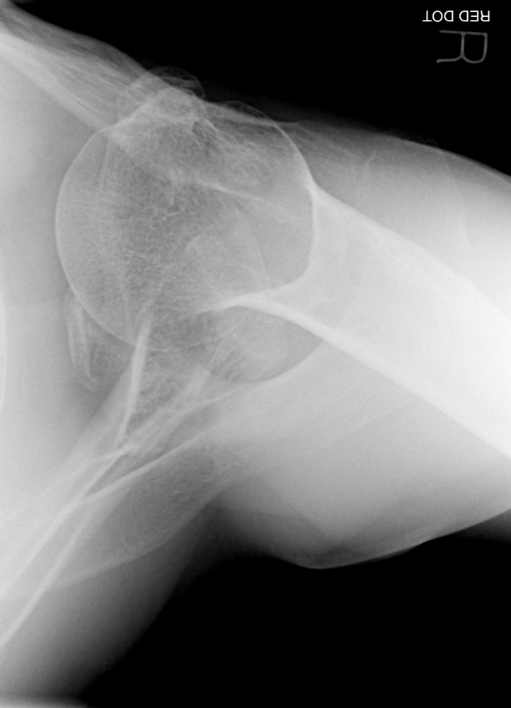
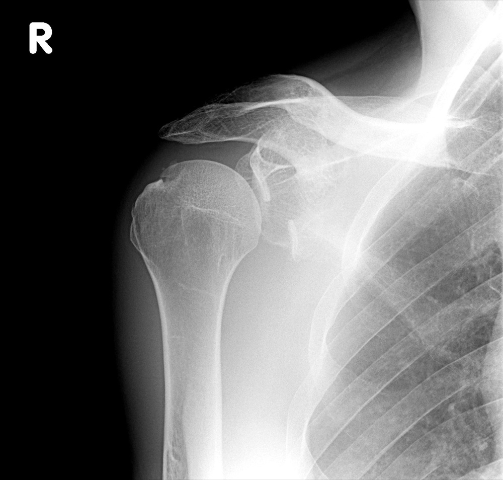
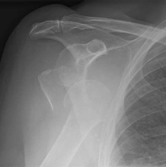
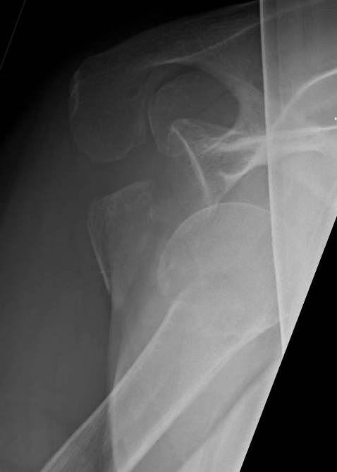
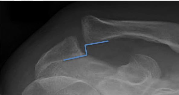
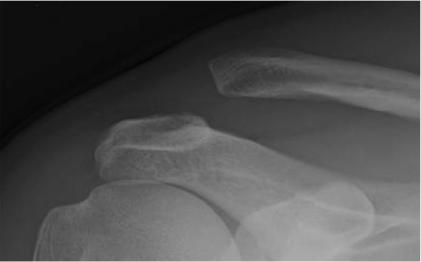
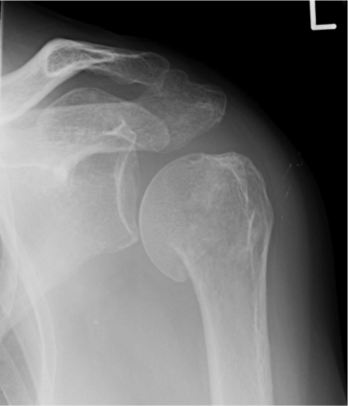
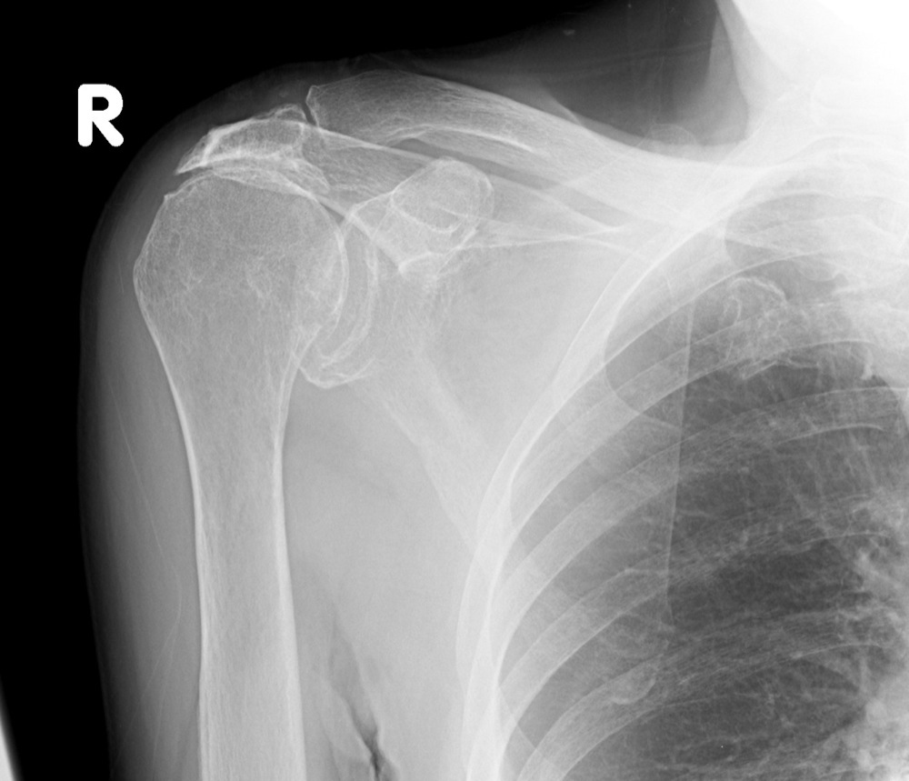
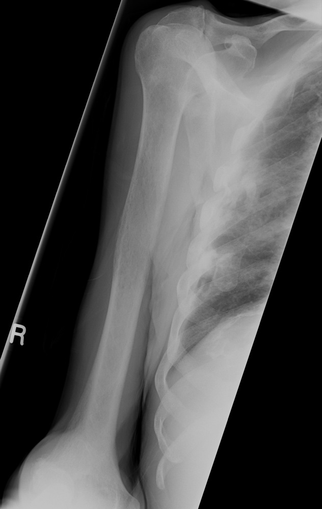
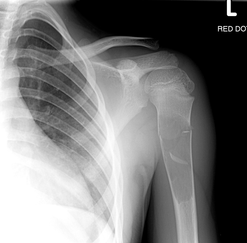
| Under 20 years and over 60 years | Fractured proximal humerus |

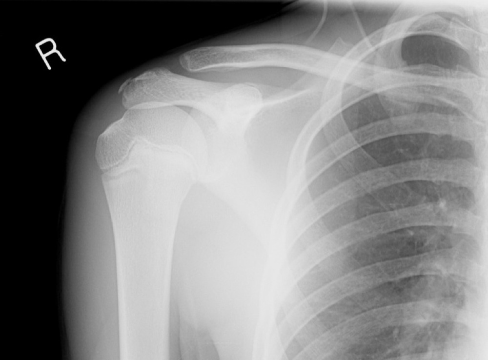
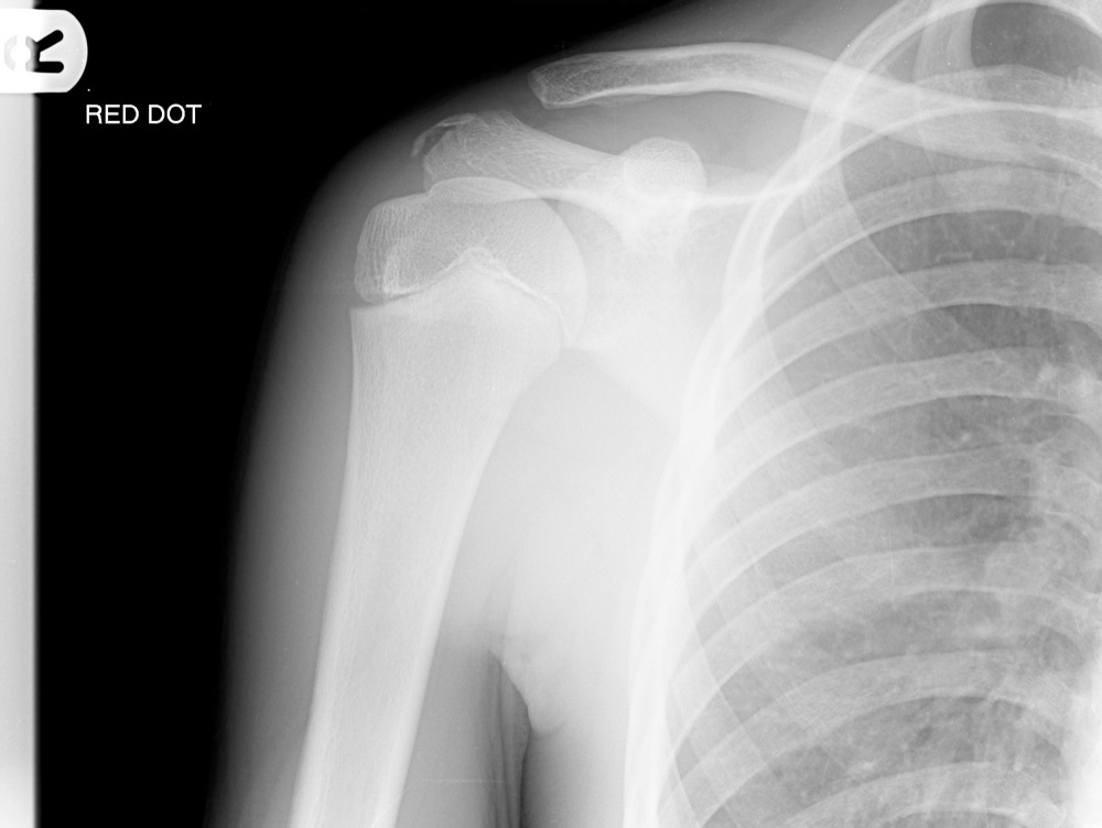
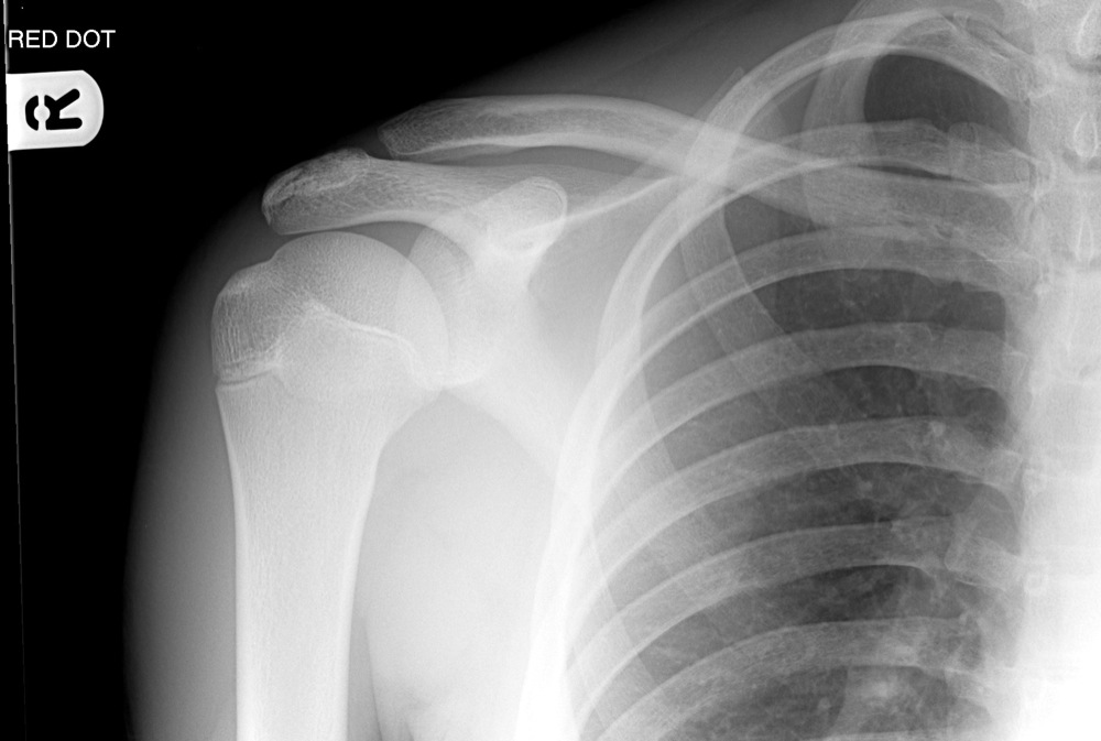
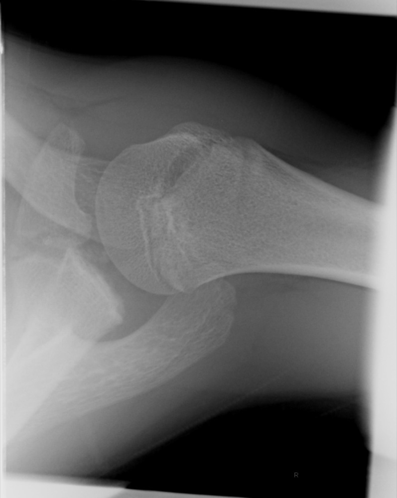
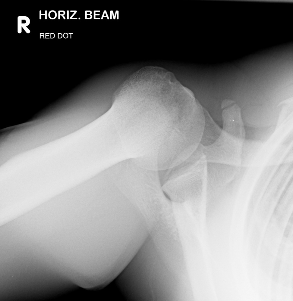
Break to the cortex
Disruption to the trabecular pattern
Sclerosis, suggesting impaction