Norwich Image Interpretation Course

Heidi Nunn (Advanced Practice Reporting Radiographer)


Bone Tumours and Benign Lytic Lesions

Factors aiding diagnosis Benign lesions Non-ossifying fibroma Solitary bone cyst Aneurysmal bone cyst Enchondroma Osteochondroma Osteoid osteoma Eosinophilic granuloma Haemangioma Fibrous dysplasia Giant cell tumour Malignant tumours Osteosarcoma Ewing's sarcoma Chondrosarcoma Metastases Multiple myeloma Osteomyelitis

(hover over images to zoom, click to enlarge)

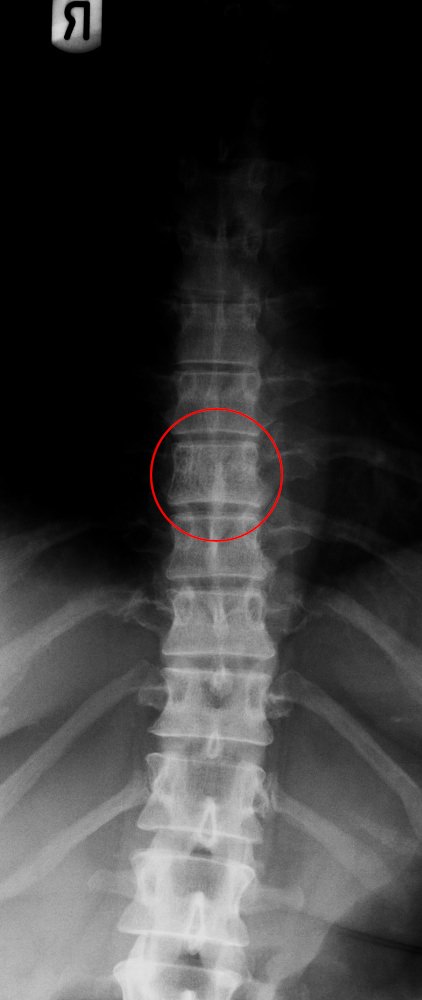
When interpreting whether an image is normal or abnormal, it is common to come across incidental lytic lesions, which, depending on their appearance, must be classified as either a normal variant, or something which warrants further investigation.

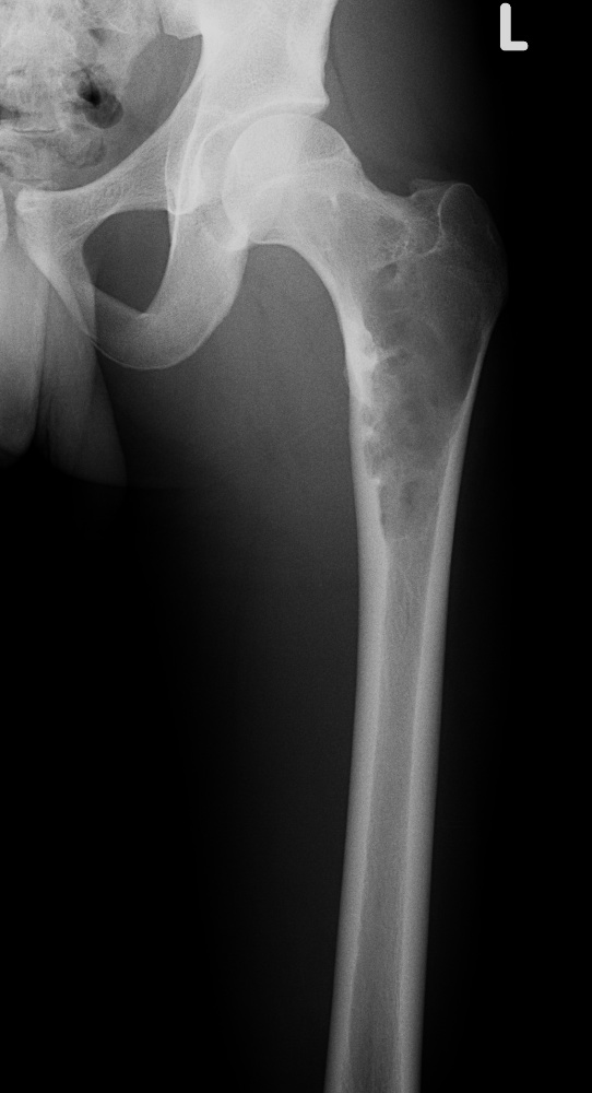
It is difficult to determine radiologically with plain radiograph imaging if a lytic lesion is benign or malignant. It is more accurate to describe whether the process looks aggressive or non-aggressive. Some factors, as outlined below, help to determine whether a lesion looks aggressive or non-aggressive, and therefore the differential diagnosis.

It is important to remember, however, that some benign processes such as osteomyelitis, can mimic malignant tumours, and some malignant lesions, such as metastases or myeloma, can look benign.

Factors aiding in the diagnosis of bone tumours and benign lytic lesions:
Age of patient
  • Specific lesions tend to occur in specific age ranges. Solitary bone cysts, non-ossifying fibromas, aneurysmal bone cysts and Ewings tumours occur in patients under the age of 30 years. Metastases and myeloma will usually occur in patients over the age of 40
Location within the bone
  • Epiphyseal, metaphyseal or diaphyseal
  • Central within the bone, eccentric or cortical
  • Lesions often arise within specific bones, and within specific areas of that bone. Giant cell tumours for example, usually arise within the distal femur or proximal tibia, and will always abut (push against) the articular surface
Size of lesion
  • Size of lesion is not necessarily an indication of how aggressive the process is, but recognition that specific lesions have a tendency to grow larger can help lead to the correct diagnosis. Solitary bone cysts within the proximal humerus, for example, often become large. A large lytic lesion is at risk of fracturing and it is therefore often prophylactically packed to prevent fracture and subsequent deformity
Monostotic (one lesion) or polyostotic (multiple lesions)
  • Multiple lesions are also not necessarily indicative of an aggressive process. Although metastases and myeloma are usually multiple, most aggressive processes demonstrate a single lesion. Similarly, benign enchondromas often become multiple within the phalanges
Zone of transition from normal to abnormal bone
  • This is often the best indicator as to whether a lesion is aggressive or non-aggressive. A very definite, sharp, and therefore narrow area (zone) between the normal and abnormal bone indicates a non-aggressive lesion. A wide, hazy, and undefined zone of transition suggests a more aggressive process. However, be aware that some benign processes (osteomyelitis) have a wide zone of transition as they are fast acting
Reactive sclerosis
  • If there is a sclerotic margin to the lesion, it is most likely non-aggressive
Pattern of bone destruction
  • Geographic = Well defined margin; non-aggressive lesion
  • Moth-eaten = Less defined margin
  • Permeative = Poorly demarcated with multiple small irregular holes. Suggests aggressive process
Presence of visible tumour matrix
  • Cartilage = Chondroid calcifications
  • Osteoid = Sclerotic
  • Fibrous = "Ground glass", hazy opacification
Host (bone) response
  • Cortical thinning, expansion and penetration. Cortical destruction suggests an aggressive process. Be aware, however, that what may appear to be cortical destruction may actually be cortical bone replacement by a fibrous or chondroid matrix, which is non-calcified and may be located within a benign lesion. This gives the false impression of cortical destruction when it is actually cortical replacement. Aneurysmal bone cysts, for example, often cause such thinning of the cortex as to make it undetectable radiographically
Periosteal reaction
  • Periosteal reaction will occur whenever the periosteum is irritated. This may be due to a malignant process, a benign lytic lesion, osteomyelitis, or trauma. The appearance of the periostitis will give an indication as to cause:
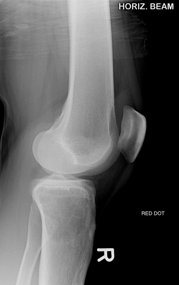
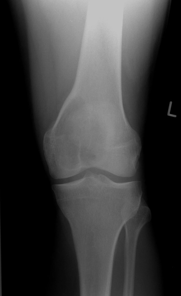
  • Benign periostitis looks thick, wavy, dense and uniform, as it is slow growing and therefore gives the periosteum time to lay down new bone
  • Aggressive periostitis is often described as lamellated (onion-skinned), amorphous and sunburst as the periosteum does not have time to consolidate
Soft tissue involvement
  • Aggressive lesions often lead to cortical breakthrough to create a soft tissue mass
BENIGN LYTIC LESIONS
Non-Ossifying Fibroma / Fibrous Cortical Defect
  • One of the most common benign lytic lesions seen
  • Asymptomatic and usually an incidental finding
  • Most often seen around the knee and distal tibia
  • Non-Ossifying fibroma generally bigger than 2cm
  • Fibrous Cortical Defect generally smaller than 2cm
  • Arises in the under 30 year age group
  • Develops from cortex of metaphysis; is eccentric within the bone
  • Usually has thin, sclerotic border that is often scalloped and slightly expansile
  • Become sclerotic as healing occurs and "disappears" as it ossifies
  • Therefore not seen in the over 30 age group
Non-ossifying fibroma - distal femur    Non-ossifying fibroma - distal femur
Simple / Solitary Bone Cyst
  • Arises in the under 30 year age group
  • Begins within the physeal growth plate and extends into diaphysis
  • Centrally located within a long bone
  • Most commonly occurs in the proximal humerus
  • In the calcaneum it is triangular, and located antero-inferiorly as this is an area that does not receive stress, and therefore develops atrophy of the bony trabeculae
  • Also called unicameral bone cyst, however there is not always just one compartment
  • Asymptomatic, unless it is fractured, which often occurs
  • "Falling fragment sign": cortical fragments produced from pathological fracture, that have sunk to the bottom of the fluid filled lesion
Solitary bone cyst - proximal humerus with fracture    Solitary bone cyst - calcaneum
Aneurysmal Bone Cyst
  • Arises in the under 30 year age group
  • Presents with pain and swelling
  • Expansile lytic lesion with a thin sclerotic margin
  • Eccentrically located in the metaphysis of a long bone, adjacent to the unfused physeal growth plate
  • May be secondary to an underlying lesion, eg giant cell tumour
Aneurysmal bone cyst - proximal humerus    Aneurysmal bone cyst - proximal humerus
Enchondroma
  • Most commonly seen in the phalanges
  • Asymptomatic but commonly fractures
  • Well-defined with narrow zone of transition
  • Lobulated
  • Can become slightly expansile
  • Causes endosteal scalloping and cortical thinning
  • Ollier's Disease = Multiple enchondromas
  • Maffucci's Syndrome = Multiple enchondromas with soft tissue haemangiomas
  • Contain calcified chondroid matrix (irregular, speckled) when located away from phalanges
Enchondroma - proximal phalanx with pathological fracture    Ollier's disease with pathological fracture
  • Differential diagnosis: bone infarct. This often occurs within femur or tibia and typically demonstrates patchy sclerosis with demineralisation
Bone infarct - distal femur    Bone infarct - distal femur
Osteochondroma
  • Arises in the under 20 year age group
  • Bony growth arising from metaphysis and covered with a cartilage cap. Extension of normal bone
  • Originates at cartilage (physeal growth plate) and migrates away from it with growth of bone
  • Continues to enlarge until cessation of skeletal growth
  • Small = asymptomatic. Large = present with swelling
  • May extend from a stalk (pedunculated) or be sessile (without stalk)
  • Multiple hereditary osteochondromas = diaphyseal aclasis
Diaphyseal aclasis    Diaphyseal aclasis
Osteoid osteoma
  • Typically occurs in older children / young adults
  • Clinically presents with night pain relieved by aspirin
  • Central ovoid lucent nidus (less than 2cm) which is well-circumscribed
  • Reactive sclerosis with cortical thickening and expansion
  • Generally occur in the long bones, but can also be seen in lumbar vertebrae
Osteoid osteoma - 15 year old    Osteoid osteoma - 15 year old
Eosinophilic granuloma / Langerhan's cell histiocytosis
  • Presents in older children / young adults
  • Causes medullary bone resorption
  • Radiologically manifests as "punched-out" lytic lesions
  • Often seen in the skull. Will cause vertebra plana in the spine, usually thoracic
Eosinophilic granuloma    Eosinophilic granuloma
Haemangioma
  • Benign vascular tumour
  • Vertebral haemangioma; solitary lesion within vertebral body typically demonstrates coarse vertical trabecular pattern
  • Usually asymptomatic and incidental finding
  • However, within vertebral body occasionally causes symptoms of spinal cord compression
Haemangioma - T8    Haemangioma - T8
Fibrous Dysplasia
  • Long lesion in a long bone (often occurs in proximal femur)
  • Expansion and bone deformity
  • Lytic but becomes ground-glass in appearance as the matrix calcifies, and then becomes sclerotic
  • Asymptomatic, but can fracture
  • No periosteal reaction
  • May be single or multiple lesion in different locations
Fibrous dysplasia - femur    Fibrous dysplasia - tibia, with pathological fracture
Giant Cell Tumour
  • Epiphyses must be closed
  • Must be epiphyseal and abut the articular surface
  • Well-defined with narrow zone of transition
  • Must have a non-sclerotic margin
  • Eccentric within the bone
  • Usually occurs within the distal femur or proximal tibia
  • 15per cent become malignant based on recurrence rate or subsequent metastases
Giant cell tumour - proximal tibia    Giant cell tumour - proximal tibia

Giant cell tumour - distal femur    Giant cell tumour - distal femur
MALIGNANT BONE TUMOURS
Osteosarcoma
  • Most common malignant primary bone tumour
  • Arises in the under 30 year age group. Secondary osteosarcomas seen in the elderly (e.g. malignant degeneration of Paget's disease)
  • Presents with pain
  • Usually occurs towards end of long bone
  • Aggressive with a wide zone of transition
  • Sclerosis present from either tumour new bone or reactive sclerosis
  • Usually demonstrates cortical destruction
  • Often have "sunburst" type of periostitis
Osteosarcoma - 9 year old with sunburst periosteal reaction    Osteosarcoma - 9 year old with sunburst periosteal reaction    Osteosarcoma - 9 year old with sunburst periosteal reaction    Osteosarcoma - 9 year old with sunburst periosteal reaction
Osteosarcoma - 16 year old    Osteosarcoma - 16 year old
Ewing's sarcoma
  • Second most common malignant primary bone tumour in children
  • Arises in the under 30 year age group
  • Permeative lesion usually in diaphysis of long bone
  • Arises from medullary cavity
  • Aggressive with a wide zone of transition
  • Often have "onion-skinned" type of periostitis
Ewing's sarcoma
Chondrosarcoma
  • Seen in the over 40 year age group
  • Lytic, destructive lesion with calcified chondroid matrix that looks amorphous and irregular
  • May arise from pre-existing cartilage mass such as an enchondroma or osteochondroma
  • Characteristic "rings and arcs" or "popcorn" calcification
Chondrosarcoma - transverse processes L4
Metastatic Disease
  • May demonstrate single or multiple, lytic or sclerotic lesions
  • Can look benign or aggressive
  • When aggressive, often is described as having moth-eaten or permeative appearance
  • Difficult to ascertain origin of primary
  • Metastases from a primary renal tumour will always demonstrate lytic lesions
  • Breast primary often develops lytic metastases
  • Multiple sclerotic lesions, particularly in the pelvis (in an elderly man) will usually have prostate primary
  • Painful, and often develops pathological fracture with little trauma
  • Usually in the over 40 year age group

Metastasis - C5/C6    Metastasis - left ileum    Metastasis - phalanx
Multiple Myeloma
  • May be solitary or multiple lytic lesions (plasmacytomas)
  • Radiologically, often precedes clinical or haematological presentation of myeloma
  • NICE recommendations are whole-body MRI as first-line imaging
  • Skeletal surveys are useful if MRI is unsuitable
  • Diffuse and permeative lytic lesions
  • Usually in the over 40 year age group
Multiple myeloma - skull    Multiple myeloma - humerus    Multiple myeloma - femur    Multiple myeloma - thoracolumbar spine
OSTEOMYELITIS
  • Usually presents as an aggressive lucency with a wide zone of transition
  • However, can also be sclerotic and look non-aggressive
  • Painful
  • If occurring around a joint, the adjacent articular surface will be involved
  • Blurring of soft tissue fat planes / effusion
  • Osteopenia
  • Intramedullary destruction
  • Cortical destruction
  • Periosteal reaction
  • Bone dies (sequestrum)
  • New bone formation (involucrum)
Osteomyelitis - proximal tibia
Osteomyelitis - humerus

TAKE THE BONE TUMOURS AND BENIGN LYTIC LESIONS SELF-TEST

- return to top -