Norwich Image Interpretation Course

Heidi Nunn (Advanced Practice Reporting Radiographer)


The Elbow

The Elbow

Normal anatomy Fat pad sign Anterior humeral line Radiocapitellar line Ossification centres Radial head Coronoid process Supracondylar Lateral condyle Monteggia Avulsion medial epicondyle Pulled elbow

(hover over images to zoom, click to enlarge)
Learning outcomes
  • Assess skeletal radiographs using a systematic approach
  • Use common lines which are helpful in interpreting the image
  • Describe elbow anatomy
  • Recognise and understand the significance of the fat pads
  • Understand mechanisms of injury and the likely fractures/dislocations which may result
  • Understand less common fractures around the elbow
  • Accurately describe dislocations and associated fractures
  • Understand how injury patterns differ in adults and children
  • Understand common eponyms
  • Identify normal paediatric anatomy and the development of secondary ossification centres
  • Recognise normal variants and their significance
Normal anatomy
  • Capitellum articulates with the radial head (lateral).
  • Trochlea articulates with the olecranon (medial).
  • The articulating surfaces of the distal humerus are offset anteriorly to the humeral shaft (as viewed on the lateral radiograph):
Normal elbow
The fat pad sign
  • Anterior and posterior fat pads are located within the coronoid and olecranon fossae respectively.
  • An intra-articular fracture will cause leakage of blood and marrow into the joint to give a joint effusion. This displaces the fat pads away from the bone.
  • The anterior fat pad is normally visible as a lucent line hugging the anterior humeral cortex. It is abnormal if the "sail sign" or "thorn sign" is seen (elevated anteriorly):
Normal (left), raised anterior fat pad (right)
  • A visible posterior fat pad is always abnormal, and always due to a large effusion. Not all effusions are associated with fractures, however the chances of an underlying occult fracture being present are greatly increased if an effusion (and displaced posterior fat pad) is seen. Possible underlying fracture may be an undisplaced radial head fracture or supracondylar fracture:
Normal (left), raised posterior fat pad and fracture radial neck (right)
  • Normal fat pads do not exclude a fracture. Injury to the elbow may rupture the joint capsule enabling the haemarthrosis to drain from the joint. Consequently raised fat pads won't be visualised.
Anterior humeral line
  • On a lateral projection, when a line is drawn along the anterior surface of the humeral cortex and extended, it should pass through the middle third of the capitellum (as the articulating surfaces of the distal humerus are offset anteriorly):
Normal anterior humeral line
  • This is especially useful for demonstrating subtle supracondylar fractures, as the line will pass through the anterior third of the capitellum with the characteristic posterior displacement of the distal humeral fragment (due to a FOOSH):
Abnormal anterior humeral line
Radiocapitellar line
  • When a line is drawn through the proximal radial shaft and neck, and extended through the joint, it should pass through to the articulating capitellum:
Normal radiocapitellar line
  • An abnormal radiocapitellar line will demonstrate dislocation of the radial head:
Abnormal radiocapitellar line    Abnormal radiocapitellar line
Ossification centres
  • At birth, the distal humerus and proximal radius and ulna consist of cartilage, which contain no ossification centres. Six centres of ossification progressively appear in a consistent order until the age of 12 years. The ossification centres enlarge, and fuse between the ages of 14 and 16 years (except the medial epicondyle, which fuses last at 18 to 19 years).
  • These ossification centres, and the sequence with which they appear can be remembered by the mnemonic CRITOL:
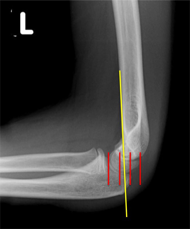
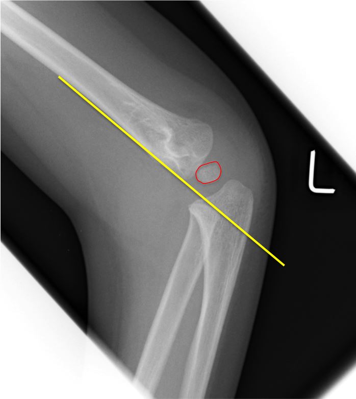
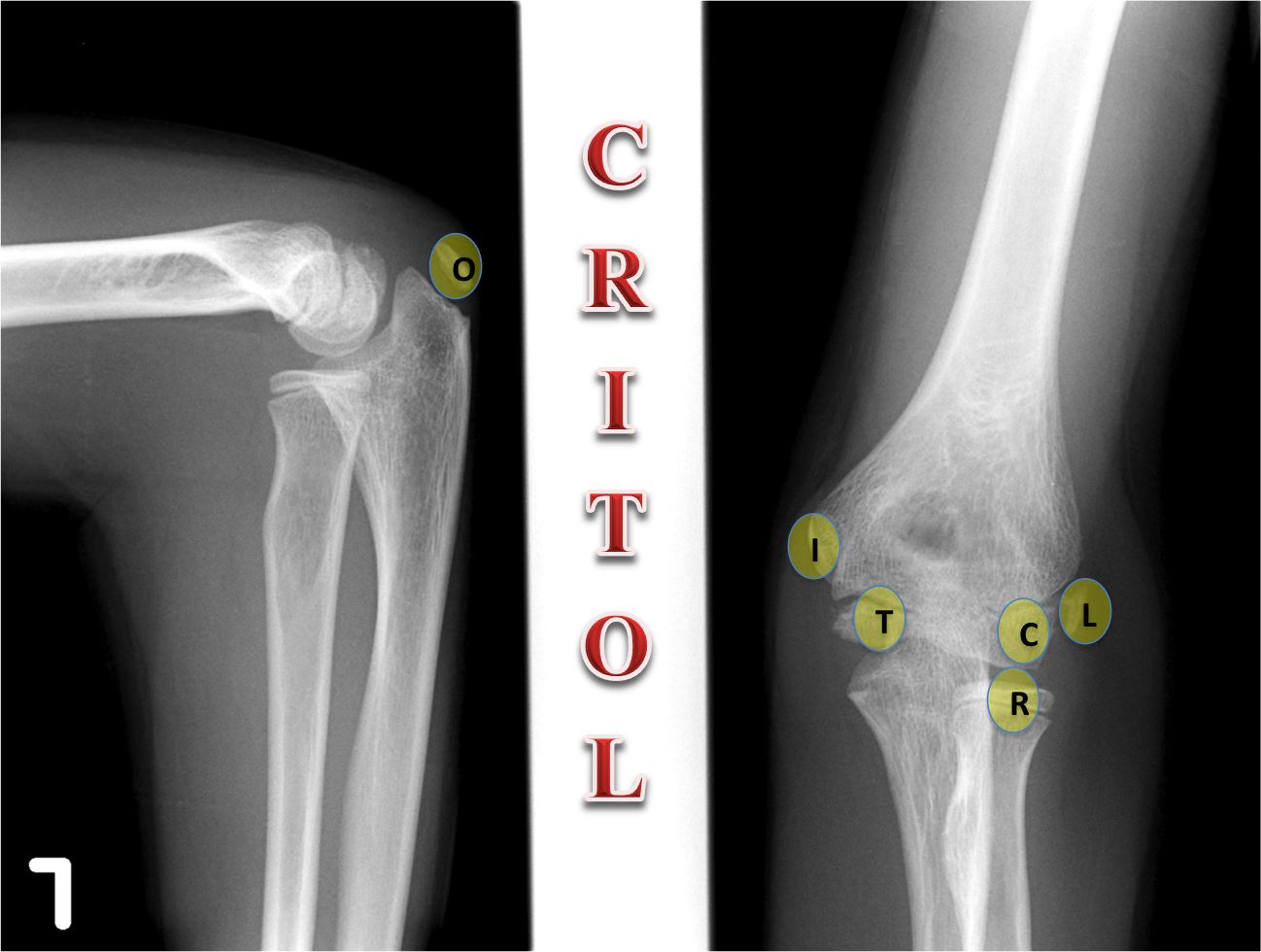
C = CAPITELLUM
R = RADIAL HEAD
I = INTERNAL EPICONDYLE (MEDIAL)
T = TROCHLEA
O = OLECRANON
L = LATERAL EPICONDYLE
  • The centres appear earlier in girls than in boys. The timing may vary, but the sequence of appearance is always the same, a fact which is invaluable when identifying internal (medial) epicondyle avulsions.
  • The apophysis for the lateral epicondyle fuses with the capitellum before fusing with the humerus.
  • The trochlea often ossifies irregularly. The apophysis for the olecranon may simulate a fracture, particularly when it is starting to fuse.
  • When assessing the paediatric elbow, always locate the ossification centres.
Radial head fracture

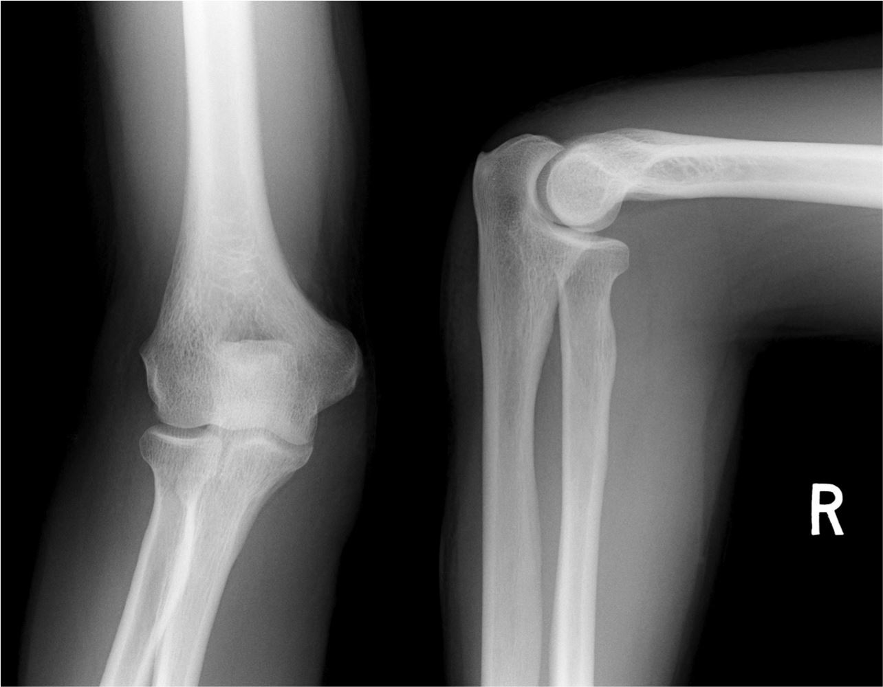
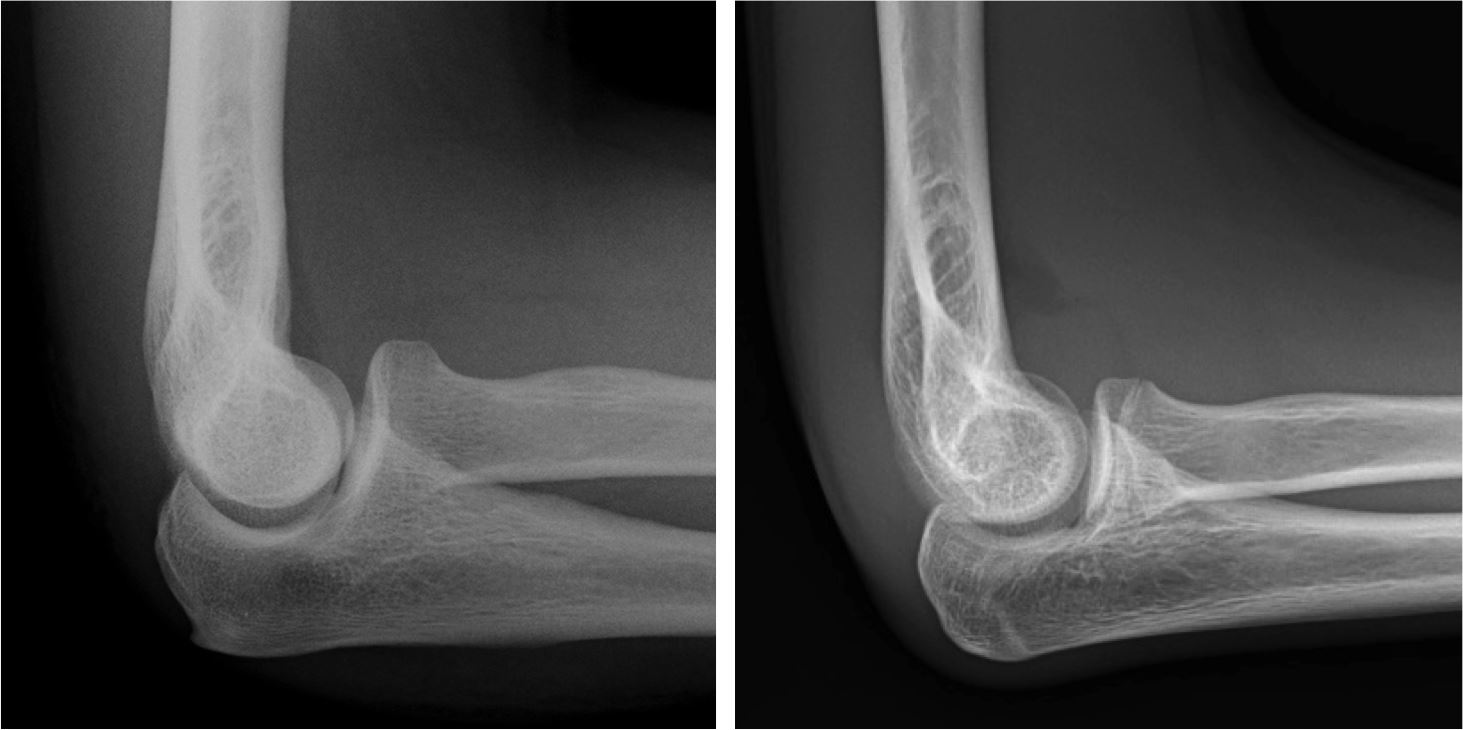
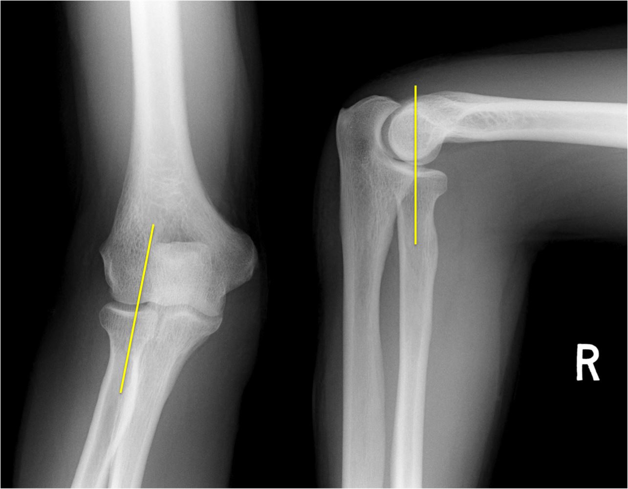
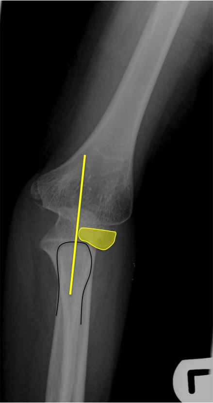
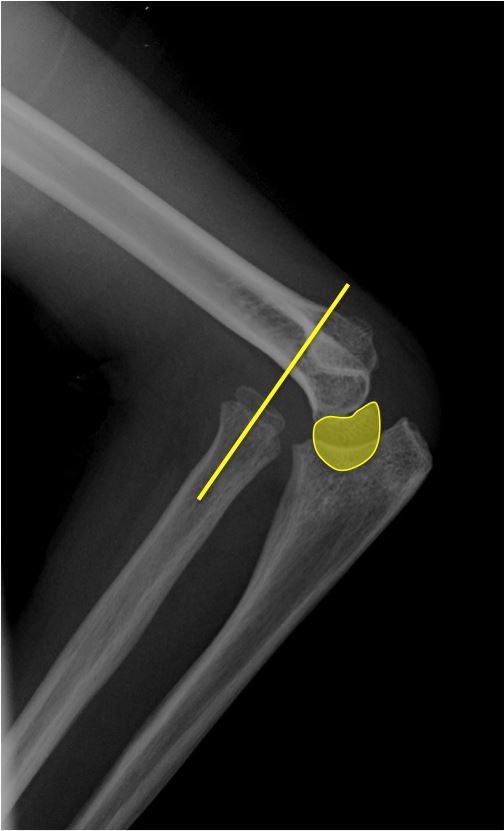
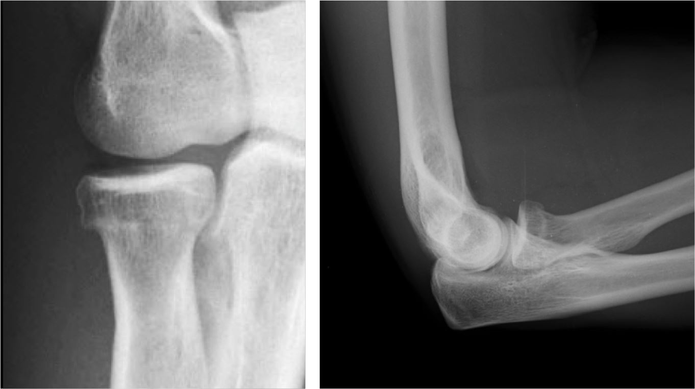
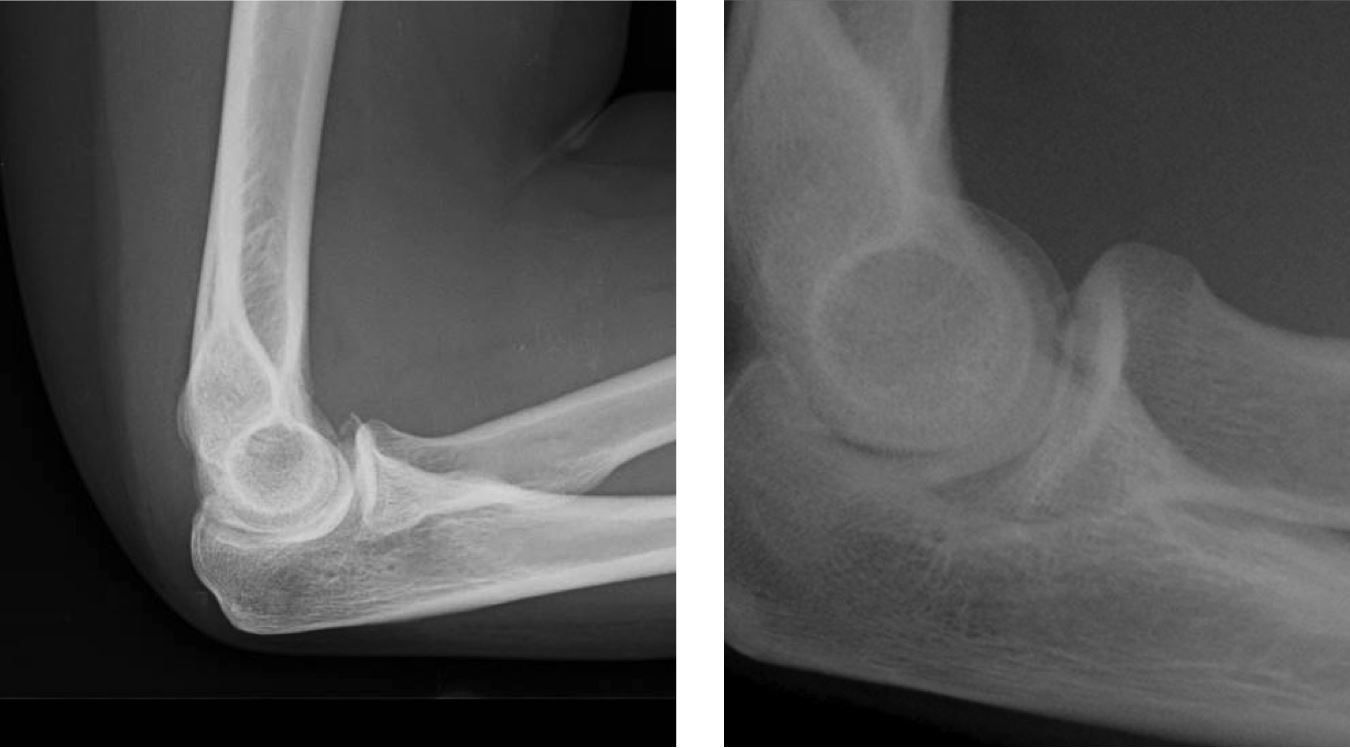
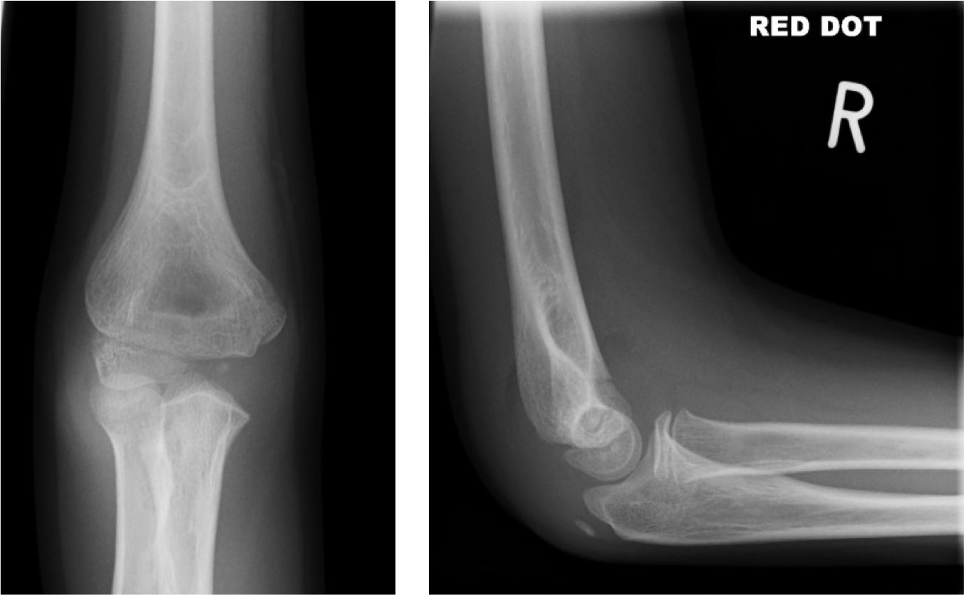
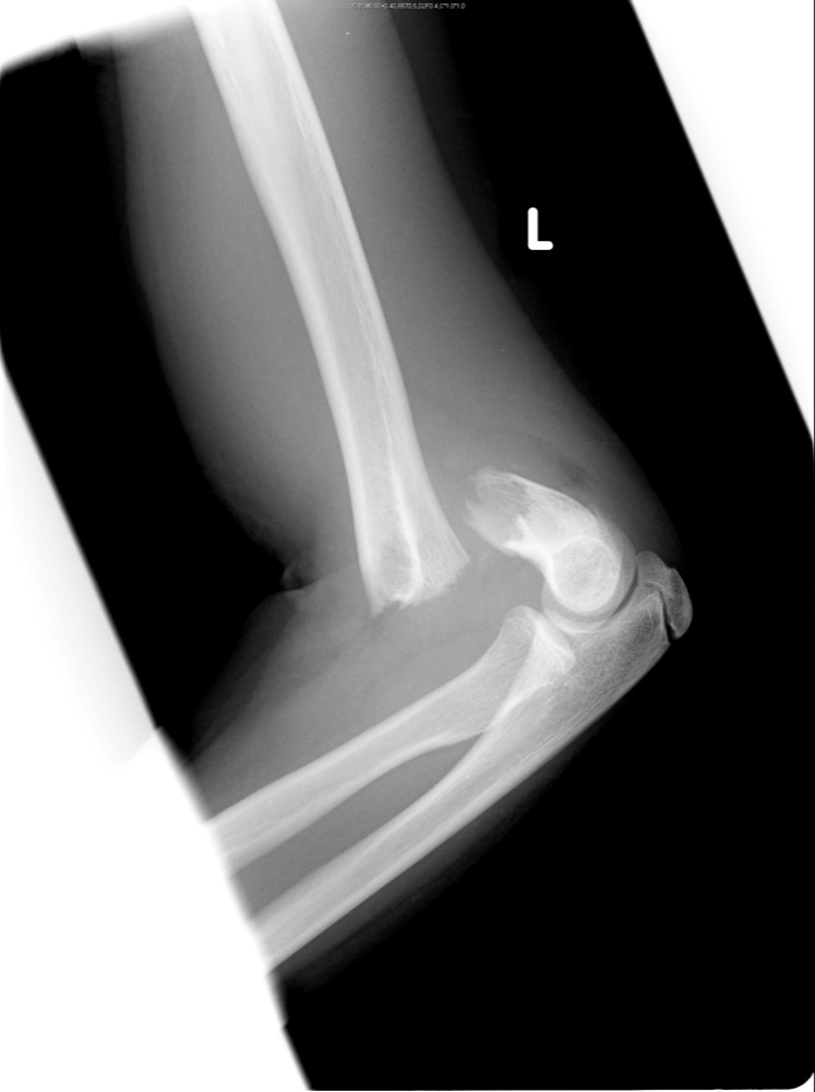
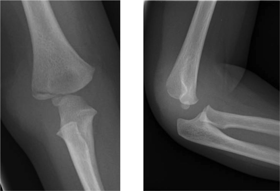
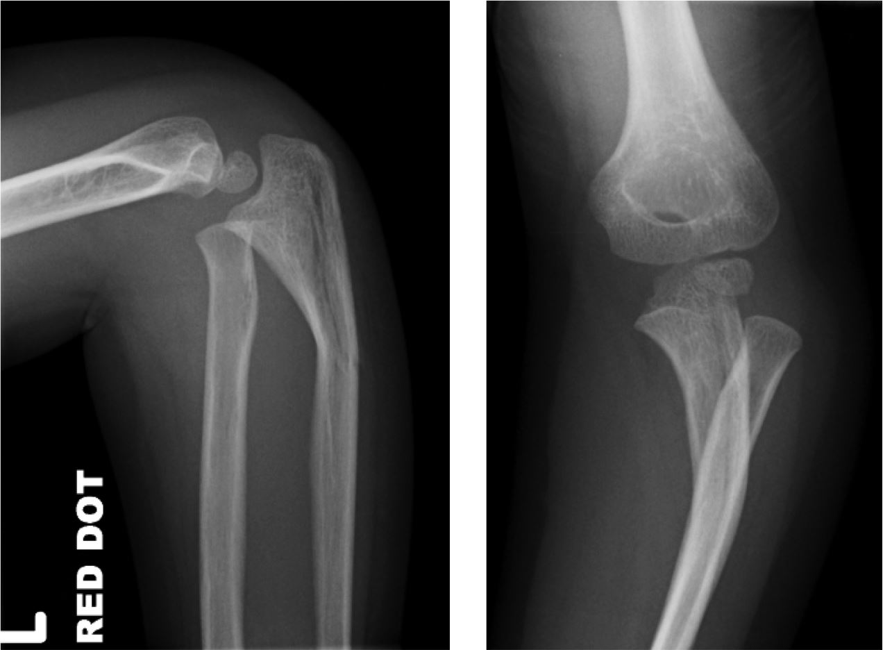
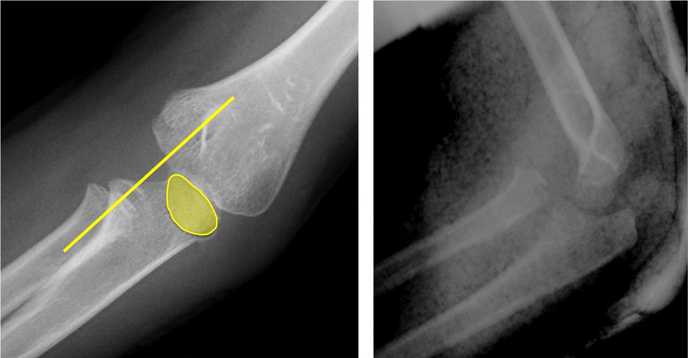
By far the most common type of fracture in adults. May be subtle; a positive fat pad sign may be the only evidence. Radial head/neck fractures may be visible as either:

  • a linear lucency extending into the joint:
Radial head linear fracture
  • a cortical break at the neck:
Radial neck fracture
Coronoid process fracture
  • Easily misinterpreted as a radial head fracture due to superimposition of the coronoid process and radial head. May be subtle if undisplaced:
Coronoid process fractures
Supracondylar fracture
  • Account for the majority of all elbow fractures in children, resulting from a FOOSH. More often than not they will have posterior displacement of the condyles and, consequently, an abnormal anterior humeral line. A positive fat pad sign is also indicative, and this may be the only clue to a subtle, non-displaced fracture:
Supracondylar fracture
  • Complications such as vascular or nerve damage arise from widely displaced fractures, as the brachial artery and median nerve both run anteriorly.
Displaced supracondylar fracture    Displaced supracondylar fracture
Lateral condyle fracture
  • Common fracture in children. It is therefore important to be familiar with the position and normal variations of the ossification centres:
Lateral condyle fracture
Monteggia fracture-dislocation
  • As the radius and ulna are a ring structure, if one displaced fracture is seen, another should be sought. The second injury may be a dislocation.
  • The Monteggia fracture-dislocation refers to a fracture of the proximal ulnar shaft with an associated dislocation of the radial head. This can be identified by drawing the radiocapitellar line.
Monteggia fracture-dislocation   Monteggia fracture-dislocation   Monteggia fracture-dislocation
  • A similar fracture-dislocation may be seen distally at the wrist (Galeazzi fracture-dislocation) where a fracture of the distal radius is accompanied by a dislocation at the distal radioulnar joint.
Avulsion of the ossification centre for the internal (medial) epicondyle
  • Although not very common, this is a significant injury in children and must not be missed.
  • The medial epicondyle is the third ossification centre to appear at the elbow and the last to fuse.
  • Injury may occur:
    1. post-dislocation.
    2. by contraction of the forearm flexor muscles that originate on the medial epicondyle. This commonly occurs whilst throwing a ball, thus avulsing the epicondyle.
  • There may be varying degrees of displacement. Minor displacement inferiorly can be more obvious than major displacement. If the ossification centre is greatly displaced, it will lie within the joint where it may be mistaken for the trochlea ossification centre. This is when knowledge of the correct sequence of appearance of the ossification centres is important and the mnemonic CRITOL is useful.
Avulsion medial epicondyle
  • If the ossification centre for the trochlea is seen but the medial epicondyle is not, an avulsion of the epicondyle must be suspected.
Pulled elbow
  • This is one of the most common elbow injuries in younger children. A sudden pull on the hand causes the radial head to be subluxed distally beneath the annular ligament, which becomes entrapped within the joint. This entrapment causes the child to hold his forearm in midpronation.
  • Treatment is simply supination of the forearm, which returns the ligament to its normal position. Radiography is not indicated.

TAKE THE ELBOW SELF-TEST

- return to top -